
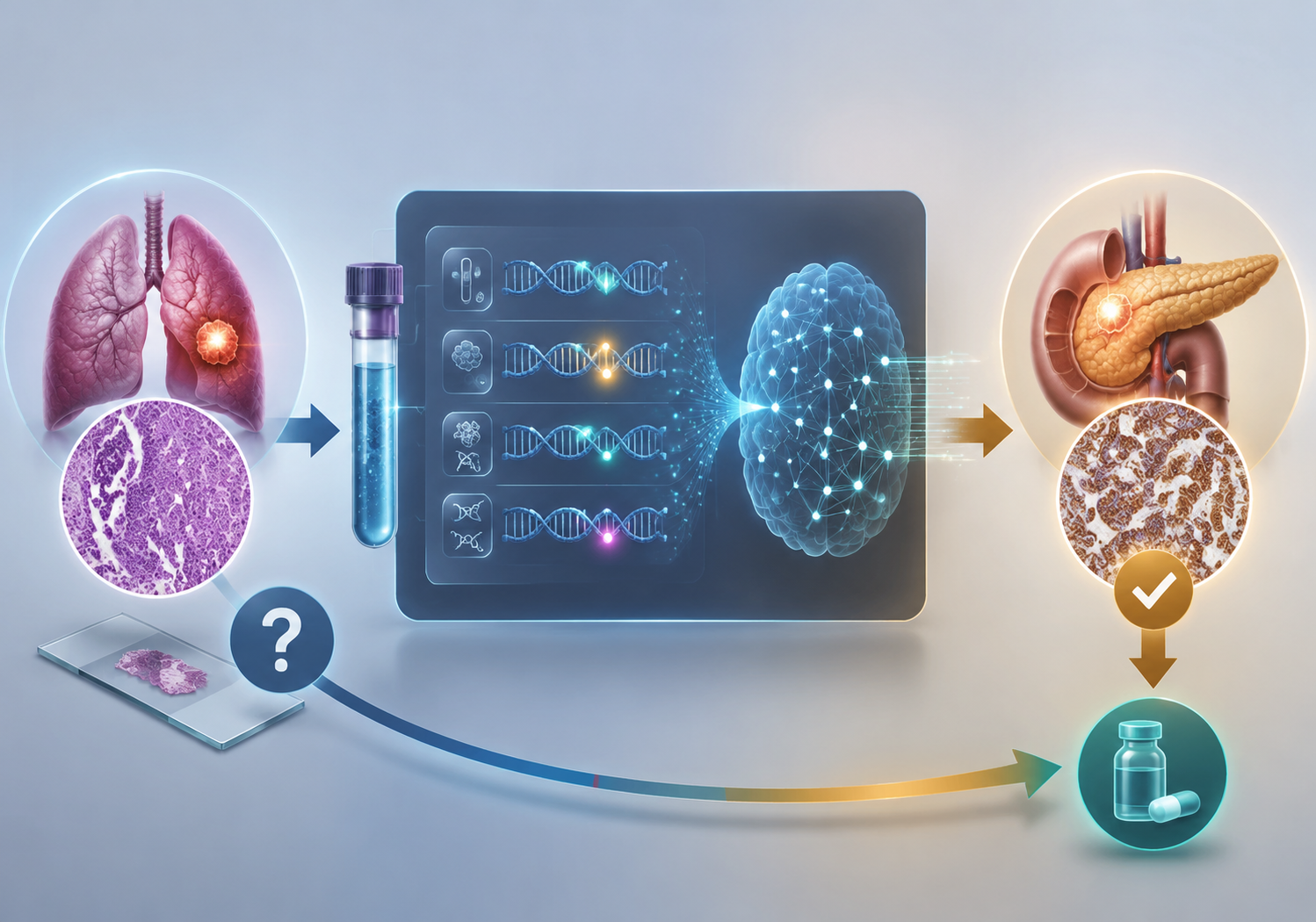
A lung cancer diagnosis can go wrong in a way that is easy to miss on paper and hard to unwind in clinic. In a 2026 JAMA Network Open study, 3.1% of 3,958 cases submitted as lung squamous cell carcinoma were actually metastases from other primary sites, and 123 cases were reclassified. [1] The useful number is not the headline accuracy of an assay; it is that the label changed for a real patient and the downstream plan changed with it.
When the label is wrong
Among the reclassified cases, 71.5% received guideline-preferred treatment changes. [1] That is the part that matters clinically: a wrong primary-site assignment can send the workup, the tumor board discussion, and the treatment choice toward the wrong disease family. The case may still look tidy in the abstract, but the chart is no longer tidy once the diagnosis has to be corrected.
AI is helping in a different part of the problem
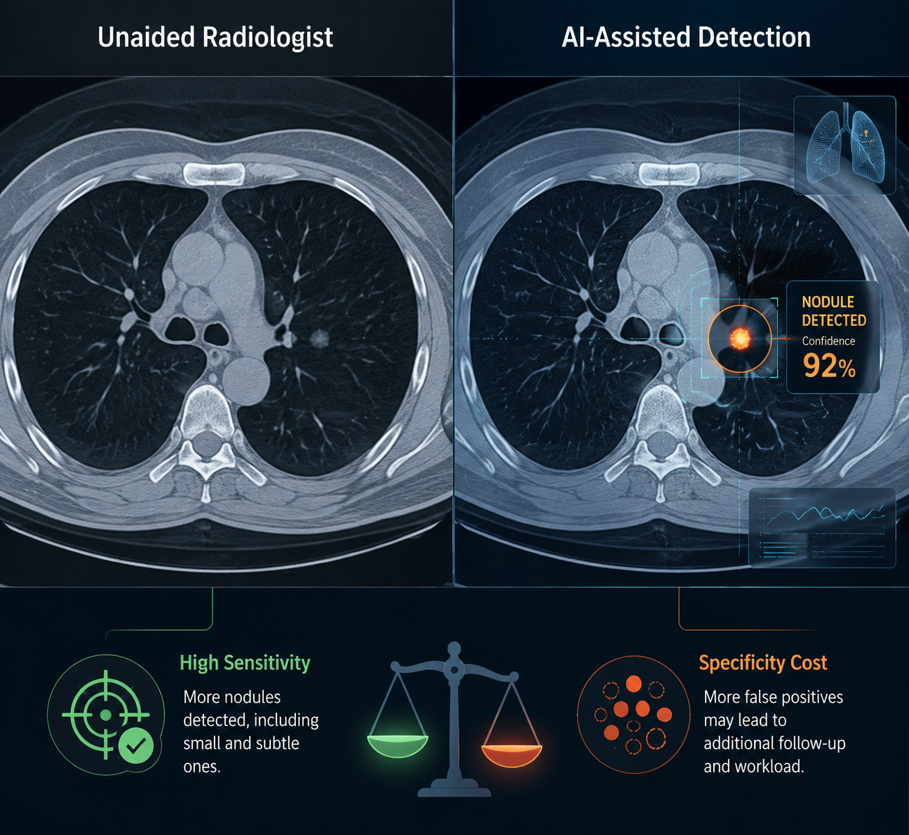
The imaging literature shows the same broad pattern from another angle. In a systematic review, AI-assisted lung nodule detection reached sensitivity of 86% to 98.1%, compared with 68% to 76% for unaided radiologists, but specificity also fell into the 77.5% to 87% range versus 87% to 91.7% for radiologist reads. [2] That is the trade-off that cannot be hidden: fewer missed nodules, but more false alarms to resolve.
That makes the 96.26% accuracy reported for the C-Swin hybrid model on the IQ-OTH/NCCD dataset interesting, but not decisive. [3] It is a single public dataset result, and it does not yet answer the question that matters in a clinic: whether the same performance holds prospectively, across sites, and in the messier case mix that real readers actually face.
Taken together, the evidence supports a careful verdict. AI can already reduce a clinically meaningful slice of lung cancer misdiagnosis, especially when molecular profiling corrects primary-site errors and imaging models recover missed nodules. [1][2][3] What is still unproven is the part that matters for routine deployment: stable specificity, prospective validation, and performance that survives contact with ordinary workflow.
References
- Primary site misclassification in lung squamous cell carcinoma with GPSai, JAMA Network Open
- AI in lung nodule detection: a systematic review, Healthcare (PMC)
- AI model boosts lung cancer detection accuracy to 96%, EMJ Reviews