
The practical question for AI in brain cancer treatment is not whether a model can produce an impressive ROC curve after the case is over. It is whether the system can help at the point where a neurosurgeon is waiting, a neuropathologist is looking at limited tissue, and the differential diagnosis changes the operation. Glioblastoma and primary central nervous system lymphoma sit uncomfortably close on imaging and sometimes on morphology, but they lead to sharply different management: glioblastoma generally pushes the team toward maximal safe resection, while PCNSL is treated primarily with chemo-radiation rather than resection.
PICTURE deserves attention because it was built for this kind of high-consequence distinction and because the peer-reviewed study behind it reports unusually strong retrospective performance. It also includes an uncertainty mechanism, a feature that matters in neuropathology because the dangerous AI system is often not the one that is occasionally uncertain; it is the one that never admits uncertainty.
| Appraisal field | Current evidence position |
|---|---|
| Tool | PICTURE |
| Clinical area | Neuropathology / CNS tumor diagnosis |
| Primary evaluated task | Distinguishing glioblastoma from primary central nervous system lymphoma on histopathology slides |
| Core study design | Retrospective multi-cohort evaluation |
| External validation | Five independent cohorts from five medical centers worldwide |
| Headline metric | 99.8% accuracy for GBM-versus-PCNSL classification on 2,141 FFPE slides |
| Regulatory status as of Q3 2026 | No FDA clearance identified; no disclosed regulatory pathway in the provided evidence |
| Risk-of-bias judgment | Promising but not deployment-ready: strong retrospective FFPE evidence, unresolved intraoperative and regulatory validation |
For clinical governance, that means PICTURE is one of the more compelling peer-reviewed AI systems in brain-tumor diagnosis, but its current evidence supports research interest and prospective validation planning, not routine clinical deployment.
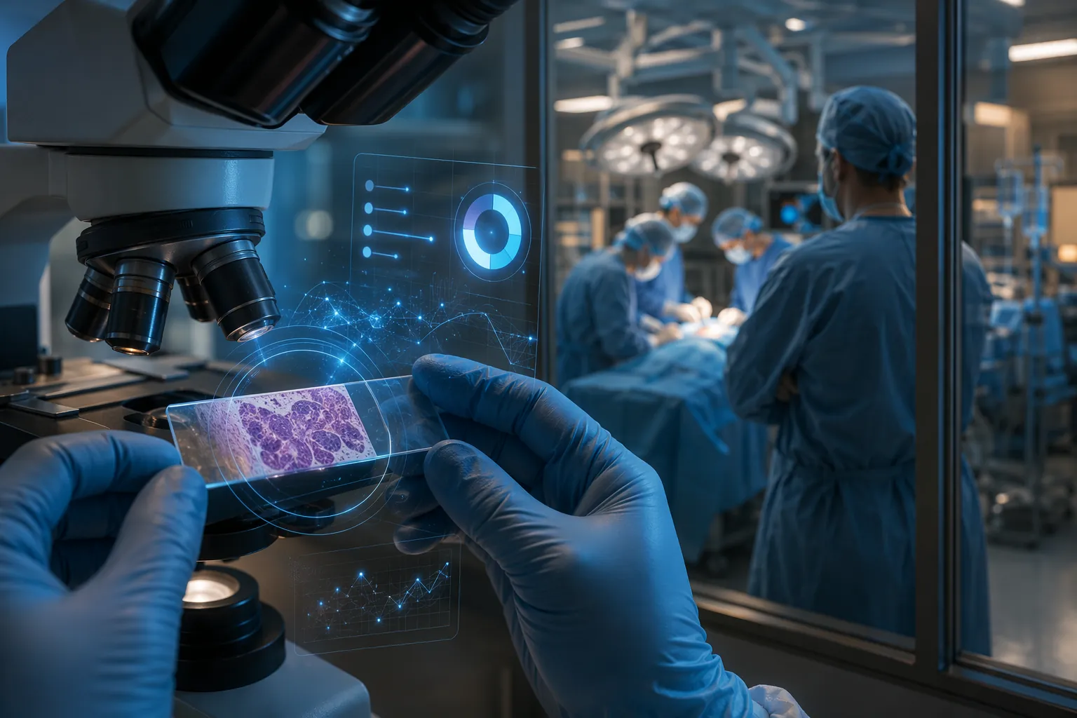
Why the 99.8% result matters, and why it is easy to overread
Yu et al. reported that PICTURE distinguished glioblastoma from PCNSL with 99.8% accuracy across five independent cohorts, using 2,141 formalin-fixed paraffin-embedded slides collected from five medical centers worldwide.[1] For a narrow differential with immediate therapeutic consequences, that is not a decorative result. It is the kind of number that properly makes neuropathologists, neuro-oncologists, and AI governance committees slow down.
The strength of the result is not only the percentage. A single-center retrospective model can look excellent because it learns the staining habits, scanner profile, tissue handling, or case mix of one institution. PICTURE’s reported performance across independent cohorts is a more serious signal. It suggests that the model was not merely recognizing one laboratory’s workflow residue.
Still, the number measures a defined task under defined conditions. The headline 99.8% accuracy comes from retrospective FFPE slides, not from a live intraoperative frozen-section workflow.[1] FFPE material is generally better processed than frozen tissue, and retrospective evaluation gives investigators control over slide selection, digitization, and timing that does not exist in the operating room. A model can be genuinely excellent on archival FFPE sections and still need a separate answer to whether it performs safely on frozen tissue when the surgical team needs an impression within minutes.
This distinction is not pedantry. In this differential, an erroneous GBM call can support more aggressive resection in a patient whose tumor may be PCNSL. An erroneous PCNSL call can reduce surgical tissue acquisition or interrupt a resection strategy in a glioblastoma case. The clinical cost is not evenly distributed across a confusion matrix; it lands in the operating room.
The human benchmark shows a real diagnostic pressure point
The PICTURE study did not simply present an AI model in isolation. It benchmarked performance against nine practicing neuropathologists. In that evaluation, the neuropathologists misclassified PCNSL as glioblastoma in 38% of test cases, and 72% of 113 misdiagnoses occurred when evaluators reported low-to-moderate diagnostic confidence.[1]
Those figures are important because they keep the appraisal anchored in actual diagnostic difficulty. The relevant claim is not that pathologists are generally poor at this differential or that AI should replace expert review. The benchmark involved nine neuropathologists and a study setting that does not fully reproduce real-world practice, where clinical history, MRI appearance, frozen-section context, immunohistochemistry, and additional tissue may affect the final interpretation. The narrower and better-supported point is that GBM versus PCNSL can be difficult even for trained readers, and that diagnostic uncertainty is not rare enough to dismiss.
That matters for governance. AI is most defensible when it is aimed at a real failure mode rather than an invented inconvenience. Here, the failure mode is visible: morphologic overlap, time pressure, and a treatment fork where the wrong impression can change what happens next.
Uncertainty is not a cosmetic feature here
PICTURE’s out-of-distribution behavior is the most interesting part of the system after the FFPE performance result. The study reports that PICTURE correctly flagged 67 rare CNS tumor types that had not been seen during training as out-of-distribution, rather than forcing them into known diagnostic classes.[1]
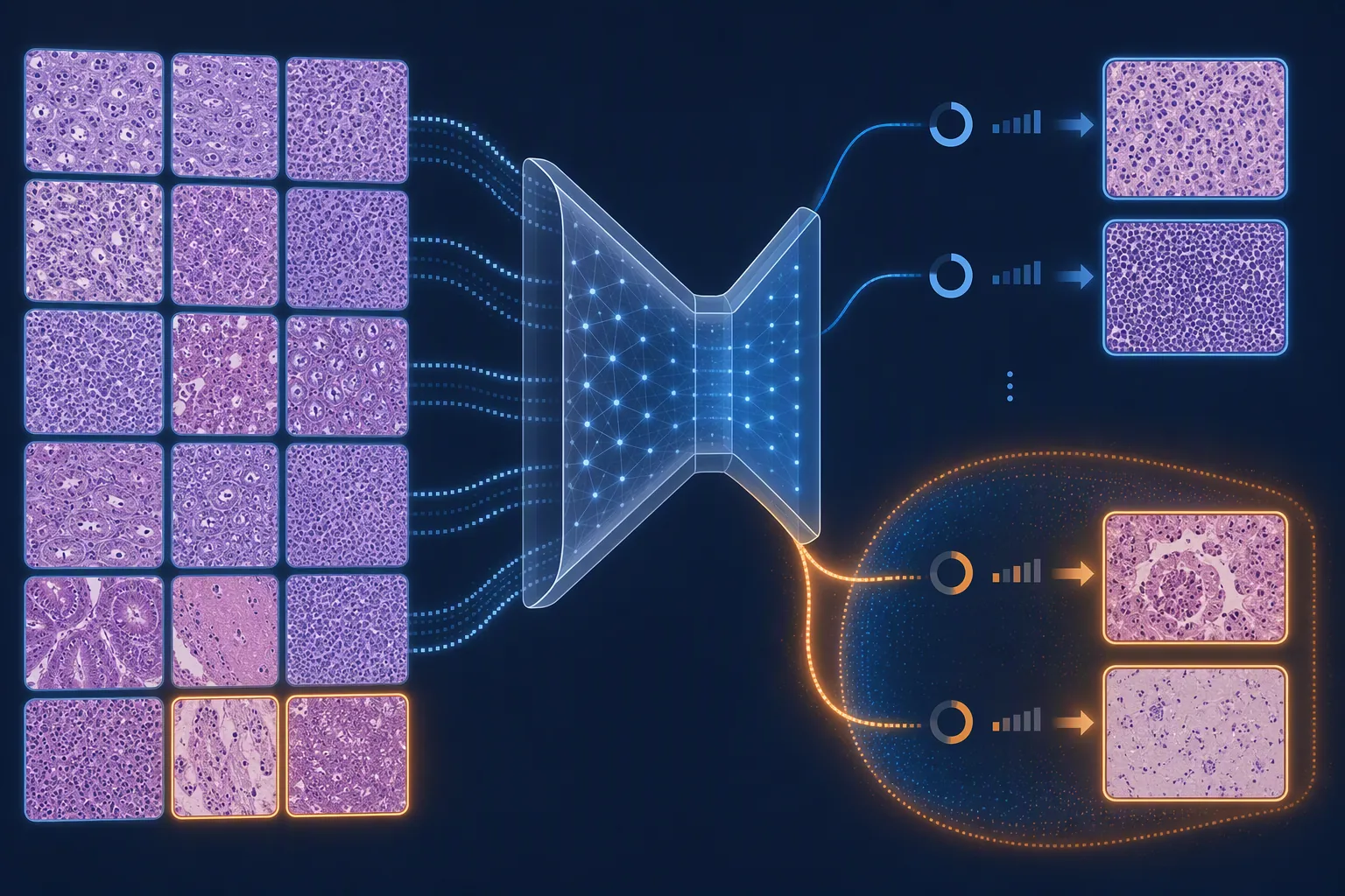
For CNS tumor diagnosis, that design choice is not a technical nicety. Rare tumors, unusual presentations, treatment effects, sampling artifacts, and nonrepresentative tissue are exactly where a closed-world classifier becomes hazardous. A forced answer can look operationally convenient while quietly converting unfamiliar biology into a familiar label. A system that can decline classification gives the pathologist and the institution a different safety posture: the model output becomes a triage signal or second reader rather than a falsely complete diagnostic authority.
The evidence still needs to be kept in its lane. Correctly flagging rare CNS tumor types in a retrospective experiment is not the same as proving safe uncertainty behavior in a live surgical workflow. In the operating room, the question is not only whether the model can detect out-of-distribution cases. It is whether the interface displays that uncertainty clearly, whether the pathologist can override or ignore it appropriately, whether the surgical team understands what an uncertain output means, and whether a delayed or withheld AI classification changes tissue handling or operative decisions.
The intraoperative gap is the decisive limitation
PICTURE is being discussed because the GBM-versus-PCNSL distinction can matter intraoperatively, but the strongest PICTURE evidence is not intraoperative evidence. The central validation result is retrospective and FFPE-based.[1] The provided evidence indicates that frozen-section performance was less characterized than FFPE performance, and no prospective intraoperative validation has been reported as of Q3 2026.[2]
That gap changes the deployment answer. Frozen sections bring different artifacts, different tissue quality, and a different time budget. The pathologist may be looking at crushed tissue, scant viable tumor, necrosis, hemorrhage, or a specimen that was not sampled where the imaging looked most diagnostic. Digital capture may need to happen quickly. The system needs to produce an output at the right moment, not after a retrospectively curated slide has been scanned and reviewed under study conditions.
A prospective intraoperative study would need to answer workflow questions that retrospective accuracy cannot answer. Which specimen is scanned? Who selects the region? How long does the model take from tissue arrival to output? Does the neuropathologist see the AI result before or after forming an impression? Are uncertain cases counted as correct refusals, workflow delays, or non-diagnostic outputs? What happens when the AI and pathologist disagree? These are not implementation details after validation; they are part of validation when the output can influence surgical behavior.
The same caution applies to downstream clinical impact. A classifier can improve diagnostic accuracy without proving that it improves patient outcomes. To support clinical deployment, PICTURE would need evidence that its use in the intended setting improves or safely supports decisions without causing unacceptable delays, overreliance, or inappropriate changes in tissue acquisition and resection strategy.
Regulatory status is not a footnote
No FDA clearance for PICTURE was identified in the provided evidence, and no regulatory pathway was disclosed.[2] For a governance committee, that is not merely a procurement inconvenience. It affects whether the tool can be used clinically, under what oversight, with what claims, and with what monitoring obligations.
The unknown predicate strategy also matters. If a developer pursues a 510(k) route, the comparison device and intended use shape the evidence package. If no adequate predicate exists, the burden may look different. Either way, a retrospective Nature Communications paper, even a strong one, is not the same thing as cleared clinical software with defined indications, locked performance specifications, quality-system controls, cybersecurity documentation, failure-mode analysis, and post-market monitoring expectations.
This is where many AI appraisals become too generous. They treat regulatory review as paperwork that follows scientific proof. In clinical diagnosis, especially in a high-stakes intraoperative context, regulatory clearance is part of the evidence boundary. It does not guarantee clinical usefulness, but its absence limits permissible clinical use and should prevent procurement language from drifting ahead of validation.
How PICTURE fits beside other intraoperative AI systems
PICTURE belongs in the broader conversation about AI-assisted brain tumor diagnosis, but it should not be forced into a simple leaderboard with systems such as SRH-plus-CNN approaches, DeepGlioma, or Sturgeon. These tools operate across different inputs and intended tasks, including H&E histology, stimulated Raman histology, and methylation-based workflows. Differences in modality, turnaround time, diagnostic target, and validation design make winner-takes-all comparison misleading.
The appropriate comparison is more practical: what tissue is available, when is the answer needed, and what decision will the output influence? PICTURE’s strongest present claim is not that it has surpassed every intraoperative AI approach. It is that, for a specific GBM-versus-PCNSL histopathology task, it has unusually strong retrospective FFPE evidence and a safety-relevant uncertainty mechanism.
What would make the evidence deployment-grade
The next evidence step is not another retrospective headline number on similar material. The needed study is prospective, intraoperative, and workflow-aware. It should test PICTURE on the specimen type and timeline where the clinical claim is being made, with prespecified handling of uncertainty and discordance.
- Prospective enrollment of surgical cases in which GBM versus PCNSL is a plausible intraoperative differential.
- Testing on intraoperative frozen-section material, not only archival FFPE slides.
- Prespecified reporting of time from tissue availability to AI output.
- Clear rules for uncertain, out-of-distribution, failed-scan, and low-quality-slide outputs.
- Measurement of pathologist-AI discordance and its effect on intraoperative communication.
- Regulatory submission or clearance aligned with the claimed clinical use.
The study would also need to avoid an easy but weak endpoint: model accuracy in isolation. If the claim is intraoperative assistance, the evaluation should show how the model behaves inside the diagnostic chain. The pathologist remains responsible for synthesis, but the AI output can still change attention, confidence, timing, or communication with the surgeon. Those effects need to be measured rather than assumed.
Current clinical appraisal
PICTURE has unusually strong peer-reviewed evidence for a narrow but clinically consequential brain-tumor differential. Its 99.8% retrospective FFPE accuracy across five independent cohorts and 2,141 slides is a serious result, and the human benchmark shows that the diagnostic problem is real rather than manufactured for an AI paper.[1] Its out-of-distribution detection is also governance-relevant because it addresses a familiar safety concern: a classifier that always chooses from known labels even when the case does not belong there.[1]
The deployment answer remains no. As of Q3 2026, the evidence does not establish that PICTURE is ready for routine clinical use in intraoperative decision-making. The missing pieces are not minor: prospective intraoperative validation, better-characterized frozen-section performance in the intended workflow, and FDA clearance or a disclosed regulatory path. PICTURE is worth watching closely and planning validation around, but the current evidence supports investigation, not adoption.
References
- PICTURE study on AI-assisted glioblastoma and primary central nervous system lymphoma diagnosis, Nature Communications, 2025.
- Reporting on the PICTURE study and clinical validation status, Inside Precision Medicine.