
The practical case for AI in diagnosis of Lewy body dementia starts before any algorithm appears. Dementia with Lewy bodies is often missed, often labeled as Alzheimer’s disease, and that error can change management in ways that matter immediately: medication choices, counseling, prognosis, driving and fall-risk discussions, and antipsychotic safety. Evidence to date supports a persistent diagnostic gap: up to 50% of DLB patients are initially misdiagnosed with Alzheimer’s disease, and a UK study found that only half of patients meeting diagnostic criteria actually received a DLB diagnosis.[1]
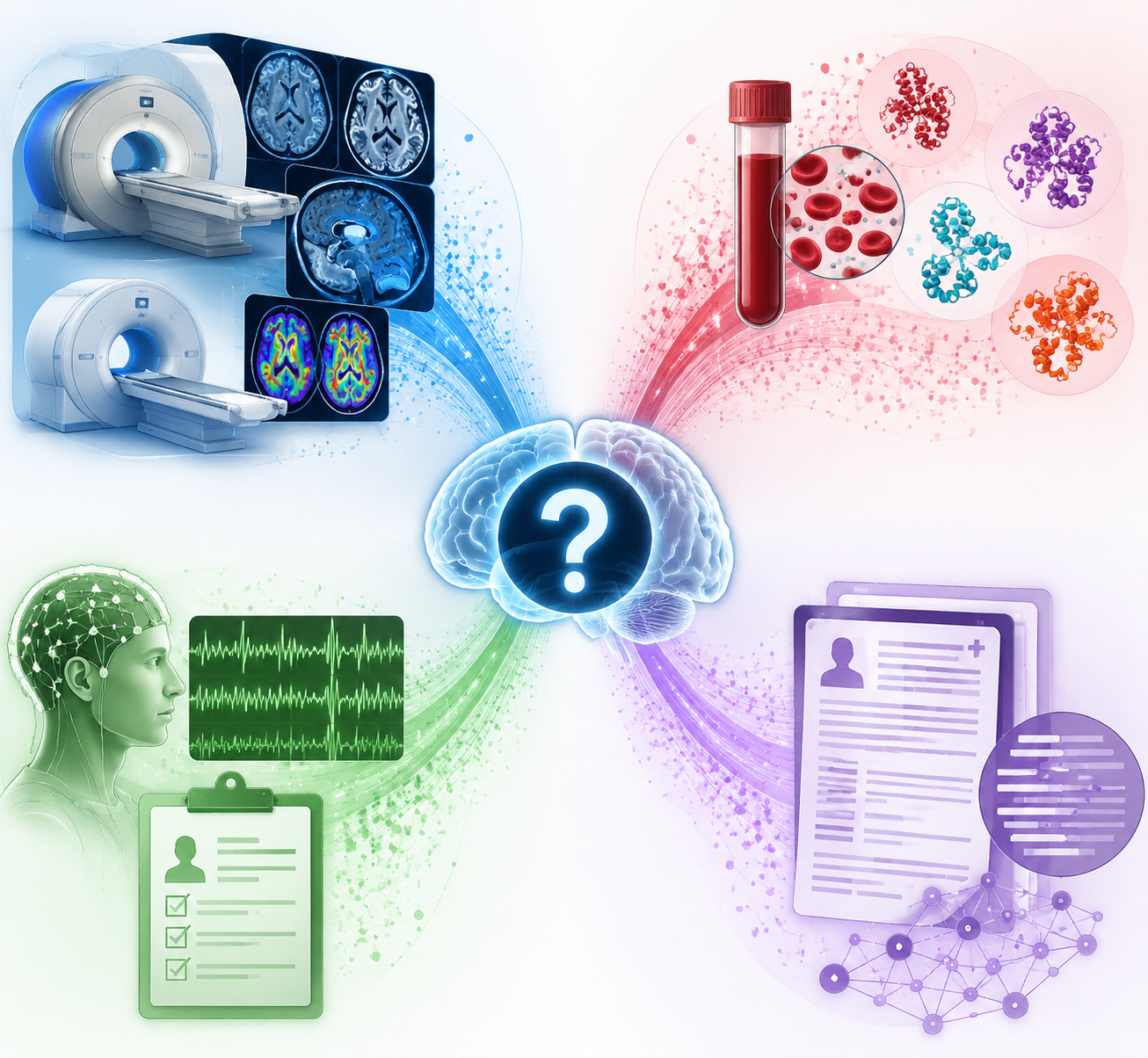
That is why DLB is a better AI target than the usual “machine beats doctor” framing suggests. The hard problem is not just separating two labels in a clean dataset. It is recognizing a disease that can present through cognitive fluctuation, parkinsonism, hallucinations, REM sleep behavior disorder, autonomic symptoms, abnormal metabolism, dopaminergic imaging, and fragments of clinical text that may never be assembled into a final diagnosis. AI is being tested against that fragmentation across several data types.
This is a modality-by-modality survey, not a product ranking and not clinical guidance. As of Q3 2026, no AI tool is FDA-cleared specifically for DLB diagnosis, and the reported performance numbers have not been tested head-to-head across the same patients, reference standards, and clinical settings. A 100% result in a small autopsy-confirmed validation set and an 88% result in a broader workflow tool are not interchangeable facts.
The Field At A Glance
| Approach | Data Modality | DLB-Relevant Finding | Validation Signal | Main Readiness Question |
|---|---|---|---|---|
| UF AIDD | Diffusion-weighted MRI | Distinguished Alzheimer’s disease from DLB with near-perfect reported accuracy | Validated at 100% on 13 autopsy-confirmed cases | How it performs prospectively and externally in larger, mixed clinical populations |
| Mayo Clinic StateViewer | FDG-PET | Identifies 9 dementia types, including DLB | Reported 88% accuracy; clinicians interpreted scans twice as fast and up to three times more accurately | Whether workflow gains translate into safer DLB diagnosis across institutions |
| 3D CNN FDG-PET work | FDG-PET | Predicted DLB, Alzheimer’s disease, and mild cognitive impairment | Included external validation | How model behavior compares with expert visual and quantitative PET interpretation |
| MUQUBIA | Multimodal MRI plus DTI | Used explainable SVM methods for neurodegenerative dementia differential diagnosis | Reported as a multimodal differential-diagnosis framework | How well DLB-specific classification holds up outside research datasets |
| Quantitative EEG ML | EEG | Differentiated DLB from Alzheimer’s disease | Prospective multicenter validation | Whether EEG can become a practical adjunct where advanced imaging is limited |
| DaT-SPECT ML scale | Dopaminergic SPECT | Tested for Alzheimer’s disease versus DLB | Real-world testing reported | Whether AI adds enough to an already DLB-relevant imaging biomarker |
| WashU blood-protein classifier | Blood proteins | 15-protein panel classified Alzheimer’s disease, DLB, FTD, and Parkinson’s disease | Tested on over 3,200 individuals; 92.3% diagnostic accuracy reported | Whether blood-based triage can reduce specialist and imaging bottlenecks without overclaiming DLB specificity |
| BU multimodal ML framework | Clinical and multimodal data | Differentiated 10 dementia types | AUC 0.96; improved neurologist diagnostic accuracy by 26% as an assistive tool | Whether assistance improves real diagnostic pathways rather than only test-set performance |
| EHR NLP | Clinical text | Found Alzheimer’s-labeled patients with documented DLB core features | 14,329 records; 18.7% of Alzheimer’s-diagnosed patients had two or more DLB core features documented | Whether NLP can trigger appropriate reassessment without creating alert fatigue |
Why Imaging Carries Much Of The Current Evidence
Imaging is the most developed part of the AI-in-DLB landscape because DLB already has clinically meaningful imaging correlates, yet those signals are not always easy to read consistently. FDG-PET patterns, diffusion changes, structural and white-matter measures, and dopaminergic imaging each ask a different question. An AI model trained on one modality is not simply another version of a model trained on another.
The University of Florida AIDD work is the sharpest example of why strong validation details matter. The tool uses diffusion-weighted MRI and was reported to distinguish Alzheimer’s disease from DLB with near-perfect accuracy. More importantly, the reported validation included 13 autopsy-confirmed cases, on which the tool achieved 100% accuracy.[2] For DLB, autopsy confirmation is not a decorative endpoint. It directly addresses the problem that clinical labels can be wrong, especially when Alzheimer’s disease is the competing diagnosis.
That same strength also defines the limit. Thirteen autopsy-confirmed cases are valuable because the reference standard is unusually strong, not because they settle general clinical performance. A model can look excellent against a small neuropathologically anchored comparison and still need prospective testing in patients whose symptoms, comorbid disease, scan quality, medication exposure, and mixed pathology resemble everyday memory-clinic practice.
FDG-PET models sit closer to an existing specialist workflow. Mayo Clinic’s StateViewer identifies nine dementia types, including DLB, and is reported to reach 88% accuracy. The workflow claim is at least as interesting as the accuracy figure: clinicians interpreted scans twice as fast and up to three times more accurately when using the system.[3] In DLB, a tool that helps clinicians see a metabolic pattern more consistently could matter even if it never replaces physician interpretation.
The reason is ordinary and clinical. A patient does not benefit from an elegant heat map unless it changes the chain of decisions: ordering the right confirmatory test, reconsidering an Alzheimer’s-only diagnosis, avoiding a risky antipsychotic, or documenting why DLB is more likely than a competing syndrome. StateViewer’s reported effect on speed and interpretive accuracy therefore belongs in the evidence map, but it should not be read as proof that DLB outcomes improve after deployment.
Other FDG-PET work reinforces the same modality cluster. Etminani and colleagues reported a 3D convolutional neural network that predicted DLB, Alzheimer’s disease, and mild cognitive impairment using FDG-PET, with external validation.[4] External validation is not glamorous, but it is one of the first filters for whether a model has learned disease-relevant signal rather than the habits of a single scanner, site, or dataset.
MRI-based multimodal work takes a broader route. MUQUBIA used an explainable support vector machine approach with multimodal MRI and diffusion tensor imaging for differential diagnosis in neurodegenerative dementia.[5] Its relevance is less that it produces a single DLB headline number and more that it reflects a plausible clinical reality: DLB diagnosis may improve when structural and white-matter information are analyzed together rather than treated as isolated impressions.
EEG and DaT-SPECT occupy different positions. Quantitative EEG machine learning has been prospectively validated across multiple centers for differentiating DLB from Alzheimer’s disease.[6] That matters because EEG is more accessible than PET in many settings and because DLB can affect network physiology in ways that routine cognitive testing may not capture. DaT-SPECT, by contrast, already has a specific relationship to dopaminergic deficit; the AI question is whether machine learning adds consistency, scale, or triage value. Chiu and colleagues reported real-world testing of a DaT-SPECT machine-learning scale for Alzheimer’s disease versus DLB.[7]
The imaging literature therefore does not point to one winner. Diffusion MRI is being used to find disease-discriminating microstructural patterns. FDG-PET models are being positioned around metabolic pattern recognition and workflow assistance. MRI/DTI approaches test multimodal anatomical signatures. EEG looks for physiological separation. DaT-SPECT asks whether an established DLB-relevant scan can be made more reproducible. The shared direction is real; the clinical role is still modality-specific.
Blood And Clinical Models Shift The Access Question
Imaging-heavy pathways are persuasive in specialty settings, but they do not fully solve the access problem. Many patients who might have DLB first appear in primary care, general neurology, psychiatry, sleep medicine, hospital medicine, or a busy memory clinic with limited advanced imaging access. Blood-based and multimodal clinical AI models are important because they could change where suspicion is raised, not just how a scan is interpreted.
Washington University researchers reported an AI-powered blood test using a 15-protein panel tested in more than 3,200 individuals. The classifier reached 92.3% diagnostic accuracy across Alzheimer’s disease, DLB, frontotemporal dementia, and Parkinson’s disease.[8] That is not the same as saying a blood draw now diagnoses DLB by itself. The supported claim is narrower and still useful: a protein classifier may help distinguish among several neurodegenerative diseases and could eventually reduce reliance on scarce specialist pathways.
The attraction of a blood panel is not only convenience. It could affect sequencing. A patient with cognitive decline and parkinsonian features might be referred earlier for DLB-focused assessment if a classifier increases suspicion. A patient with a less consistent clinical picture might be steered toward imaging, sleep evaluation, or longitudinal follow-up rather than being parked under a broad Alzheimer’s label. Those are pathway claims, not yet proven outcome claims.
Boston University’s multimodal machine-learning framework sits even closer to clinical reasoning. The system differentiated 10 dementia types with an AUC of 0.96 and improved neurologist diagnostic accuracy by 26% when used as an assistive tool.[9] The assistive design is important. DLB diagnosis often depends on reconciling symptoms, exam findings, cognitive profile, imaging, medication response, and time course. A model that improves neurologists’ decisions may be more plausible for adoption than a standalone classifier that demands trust without showing how it changes clinician judgment.
This is also where evaluation can become slippery. A model may perform well in a curated multi-dementia dataset and still face harder choices in clinic: mixed Alzheimer’s and Lewy body pathology, incomplete symptom histories, missing imaging, psychiatric comorbidity, sedating medications, or a referral note that says “memory loss” when the real clue is fluctuation. The next useful studies are not simply larger leaderboards. They need to show what information the model requires, when it is introduced, who reviews its output, and whether it changes a defensible diagnosis.
EHR NLP Is A Different Kind Of Diagnostic Tool
Natural language processing from electronic health records should not be treated as another biomarker classifier. Its value is not that it looks at a scan or protein profile and predicts DLB. It asks whether the record already contains DLB features that the final diagnosis did not absorb.
A 2025 npj Digital Medicine study analyzed 14,329 patient records and found that 18.7% of patients with an Alzheimer’s diagnosis had two or more DLB core features documented.[10] That finding is uncomfortable in the right way. It suggests that some missed DLB cases may not require a futuristic sensor to become visible. They may require a system that can notice hallucinations, fluctuations, parkinsonism, REM sleep behavior disorder, or other relevant features scattered across notes and then prompt a clinician to reassess the label.
NLP also has a different risk profile. A biomarker model can be wrong because the biology overlaps, the reference diagnosis is imperfect, or the training population does not match the patient. An EHR model can be wrong because documentation is incomplete, copied forward, negated, ambiguous, or shaped by who asked which question. The best use case may be case-finding and diagnostic audit: finding Alzheimer’s-labeled patients whose notes justify another look, rather than declaring a new diagnosis from text alone.
Why Accuracy Numbers Cannot Be Ranked Straight Across
The reported range in this field is striking: 88% for a broad FDG-PET dementia tool, 92.3% for a blood-protein classifier across several neurodegenerative diseases, AUC 0.96 for a multimodal framework, and 100% validation in 13 autopsy-confirmed cases for a diffusion MRI tool.[2][3][8][9] Those numbers are all worth attention. They should not be stacked into a league table.
- The comparison diagnoses differ: Alzheimer’s disease versus DLB is not the same task as sorting 9 or 10 dementia types.
- The reference standards differ: autopsy confirmation, clinical diagnosis, imaging-supported diagnosis, and record-based labeling carry different levels of certainty.
- The populations differ: specialty cohorts, research datasets, real-world scans, and broad EHR samples do not contain the same disease mix.
- The intended use differs: scan interpretation, differential diagnosis support, blood-based classification, case-finding, and workflow acceleration are not the same clinical job.
- The validation stage differs: external validation, prospective multicenter testing, autopsy-confirmed validation, and institutional performance reports answer different questions.
This distinction is not pedantry. A neurologist deciding whether to trust an AI suggestion needs to know whether the model has seen patients like the one in front of them. A radiologist needs to know whether a PET tool improves interpretation beyond existing quantitative and visual methods. A researcher needs to know whether the model is detecting DLB or detecting dataset artifacts correlated with a DLB label. A patient needs the diagnostic pathway to become safer, not merely more computational.
What Clinical Readiness Would Need To Show
The field has moved beyond a single experimental idea. AI approaches are now appearing across diffusion MRI, FDG-PET, multimodal MRI and DTI, EEG, DaT-SPECT, blood proteins, clinical multimodal frameworks, and EHR text. That convergence matters because DLB itself is multimodal in practice. The diagnosis rarely rests on one clue.
Readiness, however, should be judged by pathway impact. A clinically mature tool would need to show more than a strong retrospective accuracy figure. It would need larger DLB-specific samples, external validation across sites, prospective evaluation, clear comparison with specialist interpretation, calibration across disease stages, and transparent handling of mixed or uncertain diagnoses. It would also need to identify its intended decision point: triage, second reader, differential diagnosis support, record screening, or specialist workup.
Explainability may help, but only if it clarifies rather than decorates the output. A 2024 scoping review of explainable AI for DLB described SHAP-based and related approaches in this space.[11] For a DLB tool, explanation is useful when it lets a clinician see whether the model is weighting plausible features, such as a relevant imaging region or documented clinical sign, rather than an irrelevant proxy. It does not remove the need for validation.
The best near-term role may be assistive and corrective: flagging patients whose Alzheimer’s label conflicts with documented DLB features, helping radiologists read difficult metabolic patterns, prioritizing patients for specialist evaluation, or giving neurologists a structured second opinion across multiple dementia types. Those uses match the current evidence better than a claim that AI can independently diagnose DLB across settings.
As of Q3 2026, the disciplined assessment is encouraging but unfinished. AI for DLB diagnosis has strong reported signals across several data modalities, including near-perfect performance in a small autopsy-confirmed diffusion MRI validation, workflow-oriented FDG-PET results, blood-protein classification across neurodegenerative diseases, neurologist-assistive multimodal modeling, and EHR NLP evidence that missed DLB features may already be present in clinical notes.[2][3][8][9][10] None of that yet establishes an FDA-cleared DLB-specific diagnostic product, and without head-to-head comparisons the field cannot say which approach is most ready for adoption.
References
- AI in Diagnosis of Lewy Body Dementia, ClinicalMind research brief, https://clinicalmind.ai/research-brief-ai-in-diagnosis-of-lewy-body-dementia
- AI tool could help doctors diagnose dementia more accurately, University of Florida News, June 2026, https://news.ufl.edu/2026/06/ai-tool-for-dementia/
- StateViewer, Mayo Clinic News Network, https://newsnetwork.mayoclinic.org/
- 3D CNN on FDG-PET, European Journal of Nuclear Medicine, 2022
- MUQUBIA: Explainable SVM on multimodal MRI and DTI for neurodegenerative dementia differential diagnosis, Scientific Reports, 2023
- Quantitative EEG ML classifier differentiating DLB from AD, PLOS ONE, 2022
- DaT-SPECT ML scale for AD vs DLB, Journal of Personalized Medicine, 2022
- Blood test powered by AI could transform diagnosis of dementia, Washington University School of Medicine, https://medicine.washu.edu/news/blood-test-powered-by-ai-could-transform-diagnosis-of-dementia/
- Diagnosing different forms of dementia now possible using AI, Boston University Chobanian & Avedisian School of Medicine, 2024, https://www.bumc.bu.edu/camed/news-events/articles/2024/diagnosing-different-forms-of-dementia-now-possible-using-ai/
- Electronic health record natural language processing for dementia with Lewy bodies core features, npj Digital Medicine, 2025, https://www.nature.com/articles/s41514-025-00252-x
- Explainable AI scoping review for dementia with Lewy bodies, Neurology, 2024
Comments
Join the discussion with an anonymous comment.