
For clinicians thinking about dermatitis treatment during rainy season, the hard part is often not recognizing that atopic dermatitis can fluctuate. It is deciding what to do with incomplete information between visits. A patient may have photographed one worsening patch, reported poor sleep, changed topical use, or waited because the next appointment is already scheduled. By the time the visit happens, the flare may have faded, migrated, or become a story reconstructed from memory.
Seasonal context matters here, but it should not be overstated. Recent atopic dermatitis literature links disease burden with seasonal and climate-related factors, including high humidity and thunderstorm-associated pollen rupture, and reports variation in AD severity by birth season.[1][2] That does not mean every rainy week causes a flare, or that a smartphone algorithm has been specifically validated for monsoon or thunderstorm-triggered disease. It does mean that high-humidity periods expose a familiar workflow mismatch: the skin changes more often than the clinic schedule does.
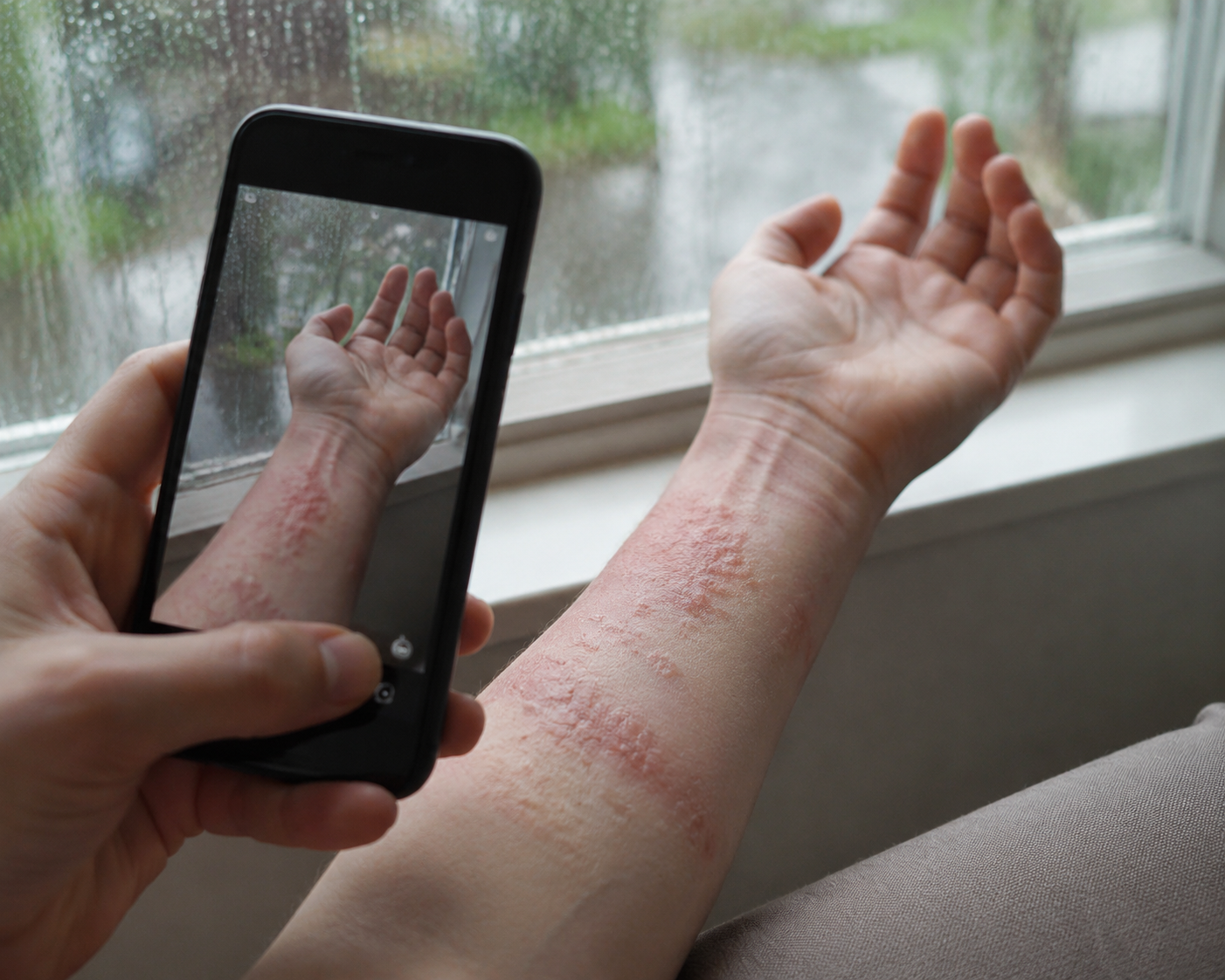
What a Smartphone Image Can Add Between Visits
A useful remote-monitoring tool does not need to solve eczema. It needs to reduce uncertainty at a specific point in care. If a patient sends a photograph during a humid stretch of worsening dermatitis, the clinician needs to know whether the visible inflammation is mild and stable, clearly escalating, or severe enough to justify faster review. A photo alone can help, but unstructured images have limits: lighting changes, body-site labeling is inconsistent, and clinicians may receive a stream of pictures without a comparable severity scale.
This is where AI-based severity scoring becomes more interesting than generic skin-photo storage. The promise is not that a phone camera can hear the whole patient story. It is that the image can be converted into a repeatable visible-severity signal, so a clinician is not comparing one remembered flare with one current patch under different lighting.
Atopiyo is one of the more concrete examples because its validation paper describes the training set, image flow, and clinical comparisons in enough detail to judge the claim. The platform was trained on 880 patient-uploaded images and validated on 220 images drawn from a larger system with more than 28,000 users and more than 57,000 images overall.[3] In that validation set, the AI reported 98% accuracy for body-part detection and 100% accuracy for identifying eczema lesions in patient smartphone photographs.[3]
The Signal That Matters: AI-TIS Versus Dermatologist Scoring
The central result is the correlation between AI-generated Three-Item Severity scores and board-certified dermatologist assessment. Across 220 validation images, AI-TIS correlated with dermatologist scores at R=0.73, with p<0.001.[3] For a smartphone-based assessment built from patient-uploaded photos, that is not a trivial finding. It suggests that the tool is measuring something clinically recognizable, not merely producing a polished consumer-app score.
| Validation measure | Reported result | Clinical interpretation |
|---|---|---|
| Training images | 880 patient-uploaded images | A modest but described image-training base |
| Validation images | 220 images | The main evidence set for performance claims |
| Body-part detection | 98% accuracy | Supports more reliable image labeling before scoring |
| Eczema lesion identification | 100% accuracy | Suggests strong lesion recognition in the validation set |
| AI-TIS vs. dermatologist scores | R=0.73, p<0.001 | Strong alignment with visible severity as judged by specialists |
| AI-TIS vs. objective-SCORAD | R=0.53 | Moderate relationship with a broader objective severity framework |
| AI-TIS vs. itch-NRS | R=0.11 | Weak relationship with patient-reported itch |
The comparison with objective-SCORAD is also important because it prevents a common overread. AI-TIS and objective-SCORAD showed a moderate correlation, R=0.53.[3] That is compatible with the idea that the algorithm captures visible disease activity, but it does not make the phone image a full substitute for a broader clinical severity assessment. Objective-SCORAD includes more than a single lesion image; Atopiyo’s analysis is focused on lesion-level image assessment.
For rainy-season monitoring, that distinction is practical. A dermatologist-correlated lesion score can help identify whether a photographed area looks worse than before, or whether visible inflammation is responding. It cannot, by itself, answer whether the disease is spreading across unphotographed areas, whether sleep has deteriorated, whether treatment adherence changed, or whether infection or another diagnosis should enter the differential.
The Itch Finding Should Change How the Tool Is Used
The weakest-looking number in the Atopiyo study may be the most clinically clarifying one. AI-TIS correlated only weakly with patient-reported itch on the itch numeric rating scale, R=0.11.[3] That result should not be hidden in a limitations paragraph. It is the dividing line between an objective digital biomarker and an eczema-monitoring system that understands patient burden.
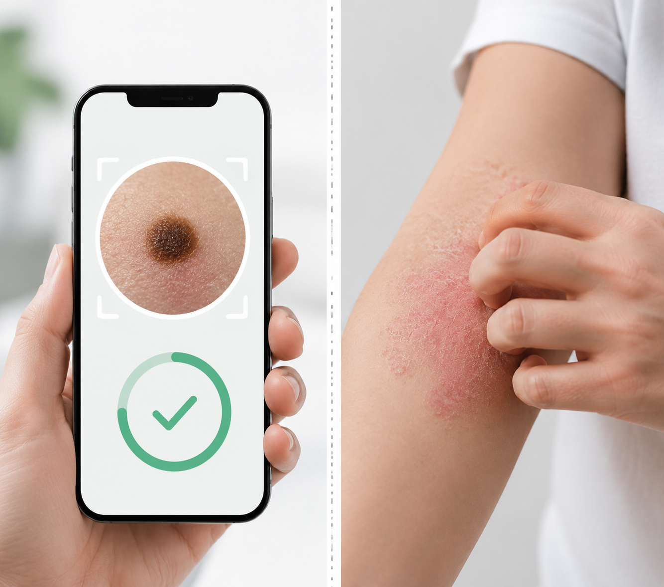
Itch often drives urgency. A patient with a visually modest lesion may be losing sleep, scratching through the night, or escalating use of nonprescribed products. Another patient may have an alarming-looking patch but little current itch. The camera can see erythema, edema, excoriation, oozing, scaling, or lesion features depending on the scoring model and image quality. It cannot feel itch.
That matters during high-humidity periods because patients are not asking for an image score; they are asking whether their disease is getting out of control. A clinician reviewing AI-TIS without itch reporting risks underestimating the patient who is visibly stable but symptomatically worse. Conversely, a high image-based severity score without symptom context may prompt unnecessary alarm if the patient is otherwise comfortable and already scheduled for follow-up.
The more defensible workflow is paired measurement: the smartphone image supplies a visible-severity signal, and the patient supplies itch, sleep disruption, pain, treatment use, and distribution changes. The AI score can make remote review more structured, but it should not be promoted as standalone triage.
Where the Evidence Is Strong Enough, and Where It Is Not
Atopiyo’s validation evidence is strongest for a narrow claim: in the studied image set, AI-generated lesion severity aligned substantially with board-certified dermatologist scoring. That is a meaningful clinical informatics result. It supports use as an objective adjunct when a clinician needs a comparable signal across repeated patient photographs.
The evidence is weaker for broader claims. The data came predominantly from Japanese users, and the developers acknowledged limited skin-tone diversity as a limitation.[3] Dermatology AI systems can perform differently when skin tone, image conditions, disease morphology, and care settings shift. A model that performs well in an East Asian user population should not be assumed to generalize across all Fitzpatrick skin types without additional validation.
The lesion-level design is another boundary. A single photographed lesion may be the right unit for tracking one stubborn flexural patch, but whole-body burden is different. If a patient photographs the worst area, AI-TIS may overrepresent global severity. If the patient photographs the easiest-to-reach area, it may underrepresent it. Remote monitoring protocols need instructions on what to photograph, when to repeat images, and how to handle new or unphotographed sites.
Regulatory status also needs plain language. No FDA clearance specific to Atopiyo was identified in the available research materials. That does not erase the validation data, but it should stop procurement-ready or autonomous-care language. In clinical settings, the tool belongs in the category of adjunctive monitoring evidence unless and until its regulatory status and intended use are clarified.
How This Fits With Broader AI Dermatology Evidence
The Atopiyo findings sit within a broader movement toward AI-supported skin assessment, but the use case is different from diagnosis. In a 2024 study, AI diagnostic support improved primary care physicians’ diagnostic agreement for skin conditions by 10%.[4] That supports the general idea that AI can improve clinician performance in dermatology-adjacent workflows, but it does not prove that every dermatology app improves management decisions, nor does it validate AD flare triage.
Severity monitoring may be the more modest and clinically useful target. Diagnosis asks the system to decide what the condition is. Flare monitoring asks whether a known condition looks better, worse, or stable over time. That narrower task is still consequential, especially when treatment decisions depend on trend, distribution, symptoms, and risk. It is also easier to integrate into existing clinician judgment than a black-box diagnostic label.
For health IT teams, the integration question is not whether the AI score is impressive in isolation. It is whether the score arrives with the photo, date, body site, patient-reported itch, current medication use, and a review pathway. A severity score that sits outside the electronic health record or patient message workflow may add another dashboard rather than reduce uncertainty.
Deployment Conditions for Rainy-Season AD Monitoring
A reasonable clinical deployment would treat AI-based image scoring as one input during higher-risk seasonal periods, not as a separate care model. The patient captures standardized images when symptoms change or at agreed intervals. The app or platform produces a visible-severity score. The patient also reports itch and other symptoms. The clinician or care team reviews the combined signal against the treatment plan and escalation criteria.
- Use AI-TIS-like scoring to compare visible lesion severity over time, especially when appointments are weeks apart.
- Require patient-reported itch and sleep impact alongside image scores, because visible severity and itch may diverge.
- Standardize image capture by body site, lighting, distance, and timing to reduce noise in repeated measurements.
- Avoid whole-body conclusions from single-lesion images unless the protocol explicitly captures representative disease distribution.
- Be cautious when applying results to populations underrepresented in the validation data, particularly patients with darker skin tones.
- Keep clinician review in the loop for treatment adjustment, urgent escalation, and diagnostic uncertainty.
The rainy-season case for smartphone AI is therefore practical, not magical. Climate and seasonal AD literature helps explain why closer monitoring may matter during humid and storm-prone periods. The Atopiyo study supplies evidence that smartphone images can generate a dermatologist-correlated visible-severity signal. The weak itch correlation supplies the necessary restraint.
Used well, AI severity assessment can help bridge the gap between intermittent visits and fluctuating disease by making remote skin changes easier to compare. Used poorly, it can turn one photographed lesion into an overconfident proxy for the patient’s whole flare. The bounded clinical position is the safer one: useful objective biomarker, not standalone triage; helpful seasonal monitoring aid, not a substitute for symptom reporting, diverse-skin validation, whole-body assessment, or clinician judgment.
References
- Season of birth and atopic dermatitis severity. PMC. 2025.
- Climate factors and atopic dermatitis flares. PMC. 2025.
- Artificial intelligence-based assessment of atopic dermatitis severity using patient-uploaded smartphone images. Allergy. May 2025.
- Artificial intelligence diagnostic support improves primary care physician diagnostic agreement for skin conditions. PMC. 2024.
Comments
Join the discussion with an anonymous comment.