
The uncomfortable timing problem in diffuse glioma surgery is that some of the information most likely to matter during resection often becomes reliable only after resection. The surgeon is already weighing functional risk against cytoreduction while the patient is open. The neuropathologist can offer an intraoperative read, but the integrated molecular diagnosis that now anchors modern glioma classification has usually depended on later testing. That mismatch is where AI in brain cancer diagnosis and research has become clinically interesting: not as a contest between algorithms and pathologists, but as an attempt to move molecular signals into the operating-room time window.
The stakes are not abstract. Malignant diffuse glioma has a median survival of about 18 months, and fewer than 10% of glioma patients are enrolled in clinical trials; one barrier is that molecular subgroup eligibility is often unavailable while surgical decisions and trial logistics are unfolding.[1] A faster molecular answer will not by itself solve survival or enrollment. It can, however, change the sequence of who knows what, and when.

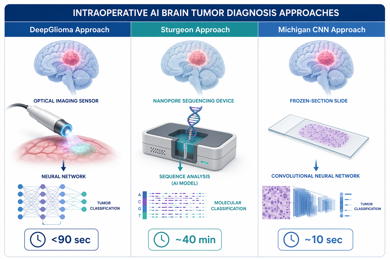
Three approaches now define the practical conversation. DeepGlioma uses stimulated Raman histology images and a deep neural network to generate molecular diagnostic predictions in under 90 seconds. Sturgeon uses nanopore sequencing and deep learning to classify tumors from sparse methylation data in an intraoperative window of roughly 40 minutes. A Michigan Medicine convolutional neural network reads frozen-section material in about 10 seconds to detect IDH-wildtype glioblastoma. Those are different inputs, different failure modes, and different deployment burdens. Treating them as one generic “AI pathology” category hides the details that decide whether an OR team can actually use them.
| System | Material analyzed | Intraoperative output window | Evidence signal | Key constraint |
|---|---|---|---|---|
| DeepGlioma | Stimulated Raman histology images | Under 90 seconds | Multicenter validation for molecular diagnosis of diffuse gliomas | Requires stimulated Raman histology hardware that is not widely available |
| Sturgeon | Sparse methylation data from nanopore sequencing | Approximately 40 minutes after sequencing begins | 72% accuracy in real-time surgical use | Misclassification risk remains clinically significant |
| Michigan Medicine CNN | Frozen-section images | Approximately 10 seconds | Institutional model for detecting IDH-wildtype glioblastoma | Generalizability beyond the single-institution setting remains unresolved |
What Changes When the Result Arrives Before Closure
A molecular call during surgery has a different meaning from a molecular call at tumor board. In the postoperative setting, the answer refines diagnosis, prognosis, adjuvant therapy planning, and trial screening. Intraoperatively, it may inform the boundary between additional resection and stopping to protect function. It may also allow a trial team to know sooner whether a patient plausibly fits a molecular subgroup, rather than waiting until the surgical episode has already passed.
That does not mean an AI output should be treated as final integrated pathology. The value is more specific: a rapid molecular signal can enter a live decision pathway, provided the team has already decided how the result will be reviewed, documented, and reconciled with standard histology, sequencing, and methylation workup. Speed only helps if the result has a defined place in the workflow.
DeepGlioma: Optical Histology Compressed Into Seconds
DeepGlioma is the cleanest example of why intraoperative AI has moved from demonstration to clinical workflow discussion. The system combines stimulated Raman histology with deep learning, allowing fresh tissue to be imaged without the same processing sequence used for conventional slides. In the reported workflow, the model generated molecular diagnostic predictions for diffuse gliomas in under 90 seconds.[1]
The important feature is not only the time stamp. DeepGlioma’s appeal is that it ties rapid optical imaging to molecular classification in a way that can occur while the surgical field is still relevant. For a diffuse glioma, that distinction matters. A diagnosis that arrives hours or days later may be scientifically richer, but it cannot help the surgeon decide whether one more pass is worth the functional risk.
The multicenter validation reported for DeepGlioma gives it more weight than a single-site feasibility story.[1] Multicenter evidence does not remove local implementation questions, but it makes the claim more serious: the model is not merely recognizing one institution’s imaging habits or one team’s sample handling. For pathology workflows, that distinction is not academic. A tool that performs only in the environment where it was built is a laboratory achievement; a tool that survives across sites begins to look like clinical infrastructure.
Its deployment constraint is equally concrete. Stimulated Raman histology is not a standard platform in most operating rooms or pathology suites. A hospital cannot adopt DeepGlioma by adding a software layer to the equipment it already uses for frozen sections. It needs access to the imaging platform, trained tissue handling, and an agreed path for neuropathology oversight. The model’s speed is therefore real, but it is tied to an instrumented workflow that many centers do not yet have.
Sturgeon: Methylation Classification Inside a Surgical Window
Sturgeon approaches the same timing problem from a different direction. Rather than using optical histology images, it uses nanopore sequencing to generate sparse methylation data, then applies deep learning to classify central nervous system tumors during surgery. Vermeulen and colleagues reported that Sturgeon could produce classifications within 40 minutes of starting nanopore sequencing.[2]
That route is mechanistically attractive because DNA methylation has become deeply embedded in CNS tumor classification. It also has an intuitive workflow appeal: obtain tissue, sequence rapidly, interpret sparse methylation data, and return a classification while the operative episode is still underway. For teams accustomed to waiting for more complete molecular workups, even a partial but reliable methylation signal could shift the conversation.
The accuracy figure forces caution. In real-time surgical use, Sturgeon achieved 72% accuracy.[2] That is not a trivial result; it shows that a methylation-based classifier can operate under intraoperative constraints. It is also not a result that supports treating the output as final. In a decision pathway where additional resection, trial pre-screening, or family counseling could follow from the label, a wrong classification has consequences.
The safest interpretation is that Sturgeon is strongest as an intraoperative classifier with an explicit uncertainty boundary. A high-confidence result may help the team prioritize next steps; a low-confidence or discordant result should not be pushed into a decision merely because it arrived quickly. The system’s value depends on how the receiving team handles confidence, non-calls, and later disagreement with standard diagnostics.
This is also where the difference between “intraoperative” and “instant” matters. Forty minutes can be clinically usable in a long brain tumor resection, but it is not the same operational category as a 10-second image classifier or a sub-90-second optical workflow. The specimen has to be obtained early enough, the sequencing has to start promptly, and the result has to return before the relevant surgical choice has passed.
The Frozen-Section CNN: Fast, Familiar Material, Narrower Evidence
The Michigan Medicine convolutional neural network sits closer to a familiar intraoperative pathology stream because it reads frozen-section images. Its reported task is narrower: detecting IDH-wildtype glioblastoma from frozen-section material in approximately 10 seconds.[1] That makes the system operationally attractive. Frozen-section workflows already exist; a rapid image model layered onto that material is easier to imagine than a new optical platform or intraoperative sequencing pipeline.
The evidence question is correspondingly sharper. A model trained and tested around one institution may perform well within that institution’s slide preparation, scanner characteristics, staining habits, tumor mix, and diagnostic conventions. Whether it travels to another hospital is a separate question. Until that is shown, the Michigan CNN is best read as a concrete institutional advance, not as a generalizable standard.
Where Molecular AI Could Alter Resection Strategy
The surgical relevance of these tools depends on whether tumor biology changes the action at the margin. In diffuse glioma, the decision to continue resection is rarely a simple matter of removing everything visible. Functional anatomy, mapping results, imaging boundaries, patient status, and the surgeon’s intraoperative impression all matter. Molecular information adds another layer: it can influence how aggressively the team interprets residual disease risk and how much weight to place on cytoreduction in that individual situation.
An intraoperative classifier does not decide the extent of resection. It can change the confidence structure around the decision. If a rapid result supports a high-grade diffuse glioma biology, the team may view additional safe resection differently than it would if the lesion appeared less aggressive. If the result is uncertain or conflicts with histologic impression, the correct response may be to avoid over-interpreting it and wait for the integrated diagnosis.
That distinction is important because “actionable” should not be inflated into “determinative.” A useful intraoperative result can narrow possibilities, support a working diagnosis, trigger additional sampling, or prompt earlier trial coordination. It does not erase the need for permanent sections, immunohistochemistry, sequencing, methylation profiling when indicated, and final neuropathology sign-out.
Trial Eligibility Is a Timing Problem Too
Clinical trial access is another place where the clock matters. Glioma studies increasingly define eligibility by molecular subgroup, but trial teams often cannot confirm those subgroups intraoperatively. With fewer than 10% of glioma patients enrolled in trials, the field does not have much room for avoidable delays in identifying potential candidates.[1]
A rapid AI-assisted molecular signal could help a research team start the right screening conversation earlier, preserve tissue with a specific protocol in mind, or flag a patient for molecular confirmation as soon as standard testing is available. That is different from enrolling a patient on an AI output alone. The credible use case is acceleration of the pathway, not bypassing the eligibility rules.
The broader research direction supports that movement toward molecularly aware classification. Work from the Indiana University School of Medicine described AI methods for glioma identification in the context of molecular classification, reflecting the larger shift away from morphology-only labels and toward integrated diagnostic categories.[3] Intraoperative systems are one practical edge of that shift: they ask whether classification can arrive early enough to matter in the procedure itself.
The Deployment Questions Are Pathology Questions
For an OR team, the key adoption questions are not limited to model accuracy. They are workflow questions with diagnostic consequences. Who selects the tissue submitted to the AI system? Does the sample represent enhancing tumor, infiltrative edge, necrosis, treatment effect, or a mixture? Is the result reviewed by a neuropathologist before it reaches the surgeon? Where is it recorded? How is it labeled if it is preliminary, probabilistic, or discordant?
- Study design: Was the system tested prospectively, retrospectively, or only in a curated dataset?
- Validation setting: Did performance hold across institutions, scanners, sample preparation methods, and patient populations?
- Platform availability: Does the hospital have stimulated Raman histology, rapid nanopore sequencing, or compatible digital frozen-section imaging?
- Uncertainty handling: Does the model provide confidence thresholds, abstentions, or rules for discordant results?
- Regulatory status: Has the system received clearance for the intended clinical use, or is it being used under a research or local validation framework?
No FDA clearance has been confirmed for DeepGlioma, Sturgeon, or the Michigan Medicine frozen-section CNN in the material reviewed for this article. That does not negate their clinical importance, but it should prevent a common category error: published performance is not the same as regulatory clearance, and local research use is not the same as broad clinical authorization.
External Validation Remains the Discipline Check
The neuro-oncology AI literature has a validation problem that cannot be ignored at the point of deployment. Khalighi and colleagues reported in npj Precision Oncology that only 29.4% of original AI neuro-oncology studies included external validation.[4] That figure is not an argument against intraoperative AI. It is a warning about how easily promising models can be over-read before they have been tested outside their development environment.
DeepGlioma’s multicenter validation is therefore not a decorative credential; it addresses one of the field’s central weaknesses. Sturgeon’s real-time surgical testing is valuable for the same reason, even though its 72% accuracy demands careful limits on use. The Michigan CNN’s speed is impressive, but its unresolved generalizability should keep it in a narrower evidence category until external performance is shown.
The most defensible adoption posture is neither enthusiasm without guardrails nor refusal to move until every uncertainty disappears. These systems are clinically meaningful because they bring molecularly relevant information into the surgical time window. Their usefulness depends on local platform availability, patient selection, neuropathology integration, prospective external validation, and clear rules for what happens when the model is uncertain or wrong. Intraoperative AI molecular diagnosis is becoming actionable, but it remains an augmenting layer in the diagnostic workflow, not the final arbiter.
References
- DeepGlioma, University of Michigan AI & Digital Health Innovation
- Ultra-fast deep-learned CNS tumour classification during surgery, Nature, 2023
- Bakas AI glioma identification, Indiana University School of Medicine, 2025
- Artificial intelligence in neuro-oncology, npj Precision Oncology, 2024
Comments
Join the discussion with an anonymous comment.