
The practical question during the 2026 Cyclospora outbreak is not whether artificial intelligence can look impressive on a conference slide. It is whether a clinical laboratory can keep finding a small, easily missed parasite when specimen volume rises, technologists are tired, and clinicians are waiting on results. For readers coming to this through the phrase using AI for detecting Cyclospora in lettuce, the first correction matters: the ARUP Laboratories deployment described here is not a produce-testing system. It is AI-enhanced microscopy for patient stool specimens, used to support clinical diagnosis during a national outbreak.
By mid-July 2026, CDC-reported figures cited in the available materials included 1,645 lab-confirmed Cyclospora cases across 34 states and 141 hospitalizations; Michigan alone had 2,640 cases, including 44 hospitalizations.[1][2] Those numbers are already large enough to stress a parasitology bench, and they still do not describe the full burden. Cyclospora reporting became optional in CDC’s Foodborne Diseases Active Surveillance Network in mid-2025, and many patients recover without testing, so confirmed case counts are a floor rather than a census.[1][2]
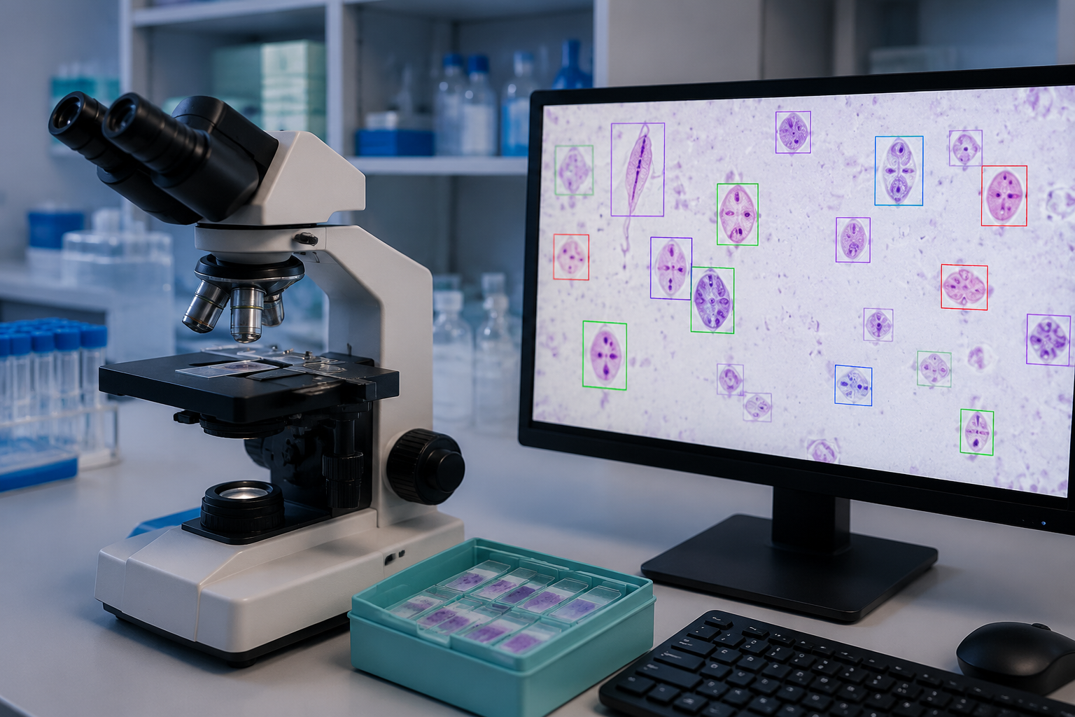
That is the setting in which ARUP’s platform becomes interesting. During the outbreak, the reference laboratory reported a roughly 200% increase in testing volume and approximately 50 positive identifications per day from specimens submitted nationwide.[2] A system that can reduce the review burden without taking the technologist out of the decision chain is not a toy in that environment. It is bench capacity.
Why Cyclospora Punishes Manual Microscopy
Cyclospora cayetanensis is not a generous organism for the microscopist. The oocysts may be shed in limited numbers, which means a slide can contain very little to find. Standard trichrome staining does not penetrate the oocyst wall, so the organism requires modified acid-fast staining for this workflow.[1][2] None of that is dramatic, but it is exactly where diagnostic misses can happen: low burden, special stain, repetitive scanning, and a surge in demand arriving at the same time.
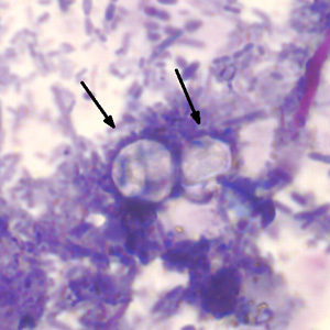
Manual review remains skilled work, not clerical work. The technologist has to know what the stain should do, what suspicious organisms look like, and when a field deserves more attention. But fatigue is not a moral failing; it is an operating condition. A bench process that depends on sustained visual concentration across a spike in specimens needs support at the exact point where attention degrades.
What ARUP Actually Deployed
ARUP’s system uses a convolutional neural network powered by Techcyte to screen digitized parasitology slides. The platform was not introduced for Cyclospora all at once. ARUP first implemented AI screening for gastrointestinal parasites on trichrome-stained slides in 2019, expanded it to Cyclospora on modified acid-fast stains in 2021, and added wet-mount screening in 2025.[1]
| Year | Workflow expansion | Why it matters |
|---|---|---|
| 2019 | AI screening for gastrointestinal parasites on trichrome-stained slides | Established the platform in routine parasite screening before the 2026 Cyclospora surge |
| 2021 | Cyclospora screening on modified acid-fast stains | Moved the tool into the stain workflow needed for Cyclospora oocyst detection |
| 2025 | Wet-mount screening added | Extended the platform across another parasitology preparation type before the outbreak year |
That timeline matters because it keeps the deployment from sounding like an outbreak improvisation. The 2026 Cyclospora workload landed on a platform that had already moved through several parasitology use cases inside one high-volume reference laboratory. That does not prove generalizability, but it does mean the tool was embedded in an existing laboratory process rather than dropped onto the bench as a one-off emergency experiment.
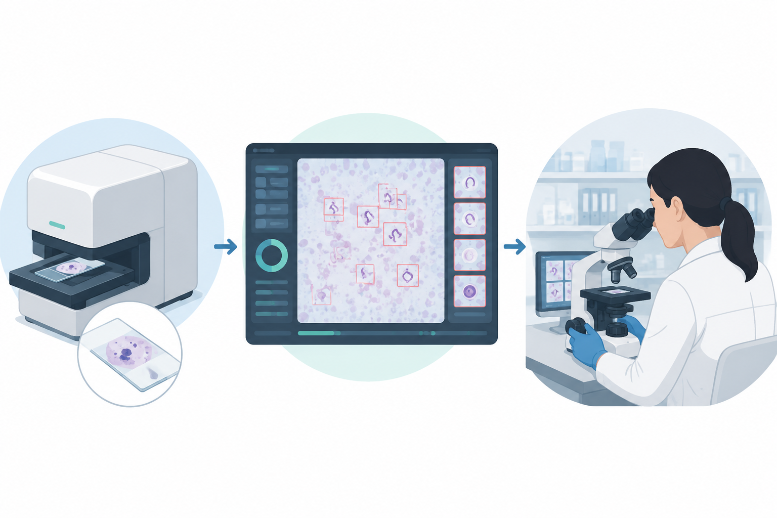
The described workflow is straightforward. A stained slide is scanned. The AI system reviews the digitized image and highlights suspicious organisms. Slides with suspicious findings are then routed to human review rather than being treated as automatically final.[1][2] In the BankInfoSecurity interview, ARUP described the AI scan-and-highlight step as taking about two minutes per slide.[2]
The important word is “highlights.” The platform is not replacing the technologist’s interpretive responsibility. It is changing where attention is spent. In a manual-only workflow, the human reviewer carries the full burden of finding the candidate organism and deciding whether it is real. In the AI-assisted workflow, the system performs an initial search over the slide image and presents suspicious areas for review. The human still has to decide what those findings mean.
The Bottleneck It Relieves
The most persuasive evidence here is operational, not decorative. A roughly 200% increase in testing volume is not an abstract “AI adoption” claim; it is a workload event.[2] In that setting, even small delays compound. More slides wait for review. More phone calls come in. More clinicians ask whether a negative result is trustworthy enough to stop looking.
A two-minute AI scan does not eliminate specimen accessioning, staining, quality control, expert confirmation, reporting, or clinical interpretation. It does, however, move a major portion of the visual search into a repeatable front-end screen.[2] That can matter most when positives are uncommon on any individual slide but common enough across a surge to keep appearing throughout the day.
The daily count reported by ARUP—about 50 positive identifications per day during the outbreak—also changes the review problem.[2] This is not a lab trying to prove that a model can recognize a few curated teaching images. It is a reference lab trying to process nationwide specimens while maintaining enough sensitivity to catch a low-burden parasite in a specialized stain.
Where the Human Review Still Sits
The system’s clinical plausibility depends on keeping human review in the chain. Suspicious organisms are flagged for technologists, not silently converted into unattended diagnoses.[1][2] That distinction is not a small regulatory or philosophical point. In parasitology, the false-negative case is the one that should make everyone uncomfortable, but false positives also consume time, trigger follow-up, and can misdirect clinicians.
The useful comparison is therefore not “AI versus people.” It is manual microscopy alone versus manual microscopy with a front-end digital search aid. In the second workflow, the reviewer is still accountable for confirmation, but the system helps decide where the reviewer’s eyes go first.
What the Evidence Supports—and What It Does Not
The available materials support a restrained positive claim: ARUP reports that validation studies showed AI-enhanced screening significantly improved sensitivity compared with human analysis alone.[1] The same deployment was associated with the lab absorbing a large outbreak-period increase in testing volume, with the slide scan-and-highlight step described at about two minutes.[2]
Those are meaningful claims, but they are not the same as a fully transparent diagnostic performance paper. The available press release and interview do not provide complete numerical sensitivity, specificity, confidence intervals, sample composition, comparator details, or subgroup performance. They tell us that sensitivity improved significantly; they do not let an outside laboratory director reconstruct the validation study or judge how the system behaved across weakly stained specimens, scant organisms, borderline artifacts, or different technologist review patterns.
| Claim | Supported by available materials | Still missing from available materials |
|---|---|---|
| The platform is deployed in clinical parasitology workflow at ARUP | Yes; ARUP describes staged implementation from 2019 through 2025 and use during the 2026 outbreak | Independent external replication |
| AI-enhanced screening improves sensitivity versus human analysis alone | Yes; ARUP reports validation studies showing significant improvement | Full sensitivity and specificity figures, validation design, and confidence intervals |
| The system improves outbreak-period throughput | Yes; ARUP reported roughly 200% higher testing volume and about 50 positives per day | How much of the throughput gain is attributable to AI versus staffing, batching, or other operational changes |
| The approach is ready for smaller laboratories | Not established | Performance data from lower-volume, differently staffed, or less digitized settings |
This is where evaluators should be careful with language. “Improved sensitivity” is not the same as “known sensitivity.” “Deployed at ARUP” is not the same as “validated everywhere.” “Aids outbreak response” is not the same as “solves Cyclospora detection.” The available evidence is enough to take the deployment seriously; it is not enough to treat it as a portable performance guarantee.
That caution is not anti-AI. It is the same standard laboratories should apply to any new diagnostic process. A method that performs well inside a large reference laboratory with specialized staining, digital scanning infrastructure, trained parasitology staff, and established review procedures may behave differently when one of those supports is weaker.
Why This Is a Clinical Workflow Story, Not a Model Story
The model type is worth naming—a convolutional neural network—but it is not the center of the clinical story. Many readers who follow computer vision AI in medical imaging already know the broad pattern: digitize an image, train a system to detect visual features, present candidate findings to a user. What separates this case from a generic imaging example is the parasitology workflow around it.
Cyclospora detection depends on the right stain, the right specimen preparation, a reviewer who knows what the organism can look like, and a reporting process that clinicians trust. The AI step sits inside that chain. If staining quality is poor, if scanning is inconsistent, if flagged findings are not reviewed by competent staff, or if the laboratory cannot manage the downstream confirmation process, the model alone cannot rescue the test.
This is also why the deployment belongs among serious AI in healthcare examples only if it is described with its constraints intact. The point is not that AI has entered parasitology. The point is that one reference laboratory appears to have used AI to strengthen a specific, fatigue-sensitive microscopy workflow under outbreak pressure.
What Smaller Laboratories Should Not Assume
ARUP’s deployment is described as a laboratory-developed test at a single reference laboratory.[2] That matters. A high-volume reference lab can justify scanners, digital workflow integration, specialized staff, validation work, and process redesign in ways that many hospital laboratories cannot. The same model, even if technically available, may not reproduce the same operational effect in a setting with lower volume, fewer experienced microscopists, different stains, slower scanning logistics, or less mature quality management.
A smaller laboratory evaluating this pattern would need answers that are not fully available in the public materials: how many true positives were included in validation; how negatives and look-alike artifacts were selected; whether performance was measured prospectively; how often the system flagged non-Cyclospora objects; how technologist review time changed; and whether sensitivity gains persisted across different operators and slide quality. Those are not academic details. They determine whether the tool reduces risk or simply moves uncertainty to another part of the bench.
The regulatory and evidence-quality context for AI for health is full of tools that look persuasive in a narrow validation setting and then become harder to judge when workflow, patient mix, or operator behavior changes. This Cyclospora case should be read with the same discipline. Its strength is that it is a real clinical deployment during a real outbreak. Its weakness is that the public evidence still leaves too much of the validation table out of view.
The Practical Judgment
ARUP’s AI-enhanced Cyclospora microscopy is a credible example of AI helping with a narrow but important laboratory problem: finding a low-burden parasite on specialized stool preparations during an outbreak surge. The reported workflow keeps technologists in the confirmation loop, uses AI to flag suspicious organisms, and appears to have helped a high-volume reference laboratory manage a major increase in testing demand.[1][2]
For now, the safest conclusion is bounded: this is proven useful enough to matter in one large reference laboratory’s Cyclospora workflow, but it is not yet proven to travel cleanly across the rest of clinical microbiology. Other laboratories should treat it as a deployable pattern to investigate, not a performance claim to inherit.
References
- Cyclosporiasis Cases Surge; ARUP’s AI-Enhanced Testing Aids — ARUP Laboratories, July 13, 2026
- AI Takes On Cyclospora Outbreak — BankInfoSecurity
Comments
Join the discussion with an anonymous comment.