
What the pooled data actually says
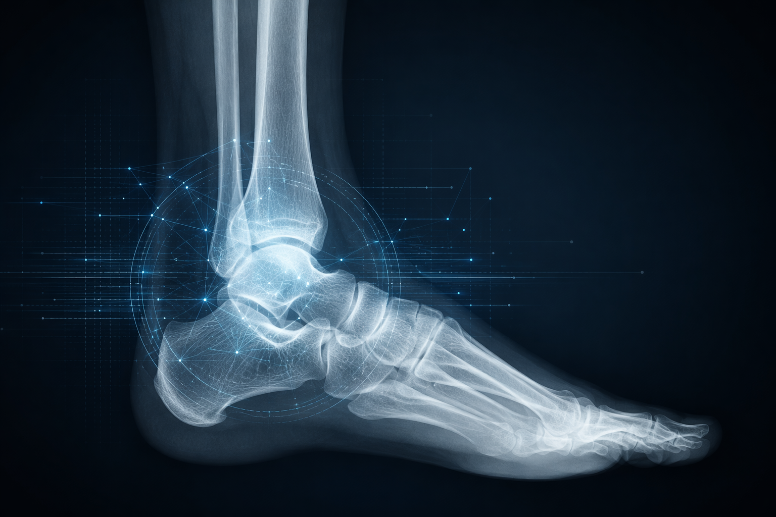
If an athlete comes in with a suspected foot or ankle fracture and the X-ray is read with AI assistance, the first question is not whether the model sounds advanced. It is whether the reader can trust it enough to avoid missing an injury that changes training, return-to-play timing, and the downstream work of unwinding a bad call. In the strongest current synthesis, a 2025 meta-analysis of 14 studies reported pooled sensitivity of 93.2% and specificity of 94.5% for AI detection of ankle and foot fractures on X-ray [1].
Those numbers are strong enough to matter in practice. They suggest the field is no longer a theoretical exercise, and they justify paying attention when an AI system is presented as a second set of eyes on a busy trauma worklist. But pooled sensitivity and specificity summarize study performance across selected datasets; they do not guarantee the same result in a local sports medicine clinic, on a different scanner, with different readers, different fracture patterns, or more difficult images.
That distinction matters because the clinical use case is not generic fracture detection in the abstract. It is foot injury diagnosis and recovery in athletes, where a subtle missed fracture can delay rehab, prolong symptoms, or shift return-to-play decisions. A model can look excellent in aggregate and still be less dependable when the anatomy is smaller, the imaging is noisier, or the patient mix is not the one the model was trained on.
Why the headline numbers still need caution
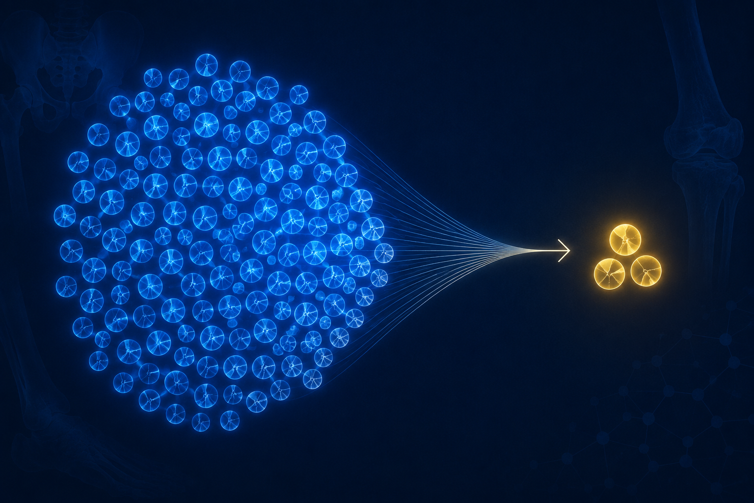
The main reason for caution is validation. In the 2023 ACFAS systematic review of 31 foot and ankle AI studies, reported AUCs ranged from 0.64 to 0.99, but only 2 of the 31 studies were externally validated [2]. That is the gap that keeps the most impressive pooled estimate from becoming a blanket clinical promise. Internal testing can show a model learned something useful from the source data; external validation is what tells a clinician whether that learning survives contact with a different patient population.
The broader fracture-detection literature points in the same direction. A separate review found that fewer than 11% of CNN fracture studies demonstrated temporal and geographic generalizability beyond a single hospital [3]. Put differently, the literature still overrepresents models that are evaluated where they were built. That does not make the results meaningless, but it does mean the average sports radiology workflow should not assume that a strong paper translates automatically into a dependable local tool.
The practical consequence is narrower than the marketing language suggests. A high pooled sensitivity can support confidence that AI is worth testing as an assistive tool, but it cannot by itself answer whether the same performance holds in pediatric athletes, in cases with casts or splints, or in centers where acquisition and reading habits differ from the study settings. That is exactly where clinicians need the evidence to be stronger than the brochure.
Commercial tools exist, but anatomy-specific performance still matters
This is not only a research conversation anymore. FDA 510(k)-cleared products including Gleamer BoneView, AZmed Rayvolve, and Imagen FractureDetect all include foot and ankle among their target anatomy [5]. That changes procurement discussions because the question is no longer whether any software can be bought; it is whether the software a department can actually deploy has been validated well enough for the cases it will see.
Clearance should not be mistaken for proof of uniform performance across every anatomy or every athletic subgroup. The foot and ankle are part of a broader fracture-detection product line for these systems, and their per-anatomy behavior may differ from the headline numbers reported across mixed datasets [5]. For a clinician reading sports trauma films, that means the relevant question is not simply whether the vendor is cleared, but whether the foot-and-ankle slice of the product has been studied in conditions resembling the local workflow.
The strongest workflow signal in the current literature is a 480-examination multi-reader study, which found that AI assistance improved reader sensitivity by 10.4% and reduced reading time by 6.3 seconds per examination [4]. That is not trivial in a busy service. It suggests the best use case may be assistive rather than autonomous: catching a fracture a tired reader might miss, or shaving a few seconds off a read without forcing a wholesale change in responsibility.
Even there, the evidence should stay in proportion. A single multi-reader study is a useful workflow signal, not proof that every site will get the same time savings or the same sensitivity gain. The effect may depend on case mix, reader experience, and how the AI output is presented in the workstation.
What the research is actually converging on
The technical approaches are also diversifying. One recent Scientific Reports study described foot fracture diagnosis using a CNN optimized by extreme learning machine, which is useful mainly as a sign that the field is still experimenting with architecture and training strategy rather than converging on a single locked-in method [6]. For clinicians, that variety is interesting only insofar as it changes what the model can do in the real world. Architectural novelty matters less than whether the system remains accurate when the image is messy and the patient is not representative of the training set.
That is also why the strongest judgment is a restrained one. Current AI systems for foot and ankle fracture detection on X-ray look promising enough to support clinical assistance, especially when the alternative is a rushed read with no second look. But as of Q3 2026, the external validation evidence is still too thin to treat those results as proven across diverse athletic populations. They are ready to be evaluated in the workflow; they are not yet ready to be assumed.
For readers interested in the broader sports-tech landscape, a companion piece on AI wearables for sports injury prediction covers a different part of the pipeline, where the signal comes from sensors rather than radiographs.
References
- Diagnostic performance of AI models for detecting foot and ankle fractures on X-ray: a systematic review and meta-analysis. PubMed, 2025. PubMed record
- Artificial intelligence in foot and ankle surgery: a systematic review. ACFAS, 2023. ACFAS article
- External validation and generalizability in CNN fracture detection studies. PMC, 2024. PMC full text
- Multi-reader study of AI-assisted fracture detection in X-ray examinations. 2024. Study record
- FDA 510(k)-cleared fracture detection tools including Gleamer BoneView, AZmed Rayvolve, and Imagen FractureDetect. U.S. Food and Drug Administration. FDA medical devices database
- Foot fracture diagnosis using a CNN optimized by extreme learning machine. Scientific Reports, 2024. Nature article
Comments
Join the discussion with an anonymous comment.