
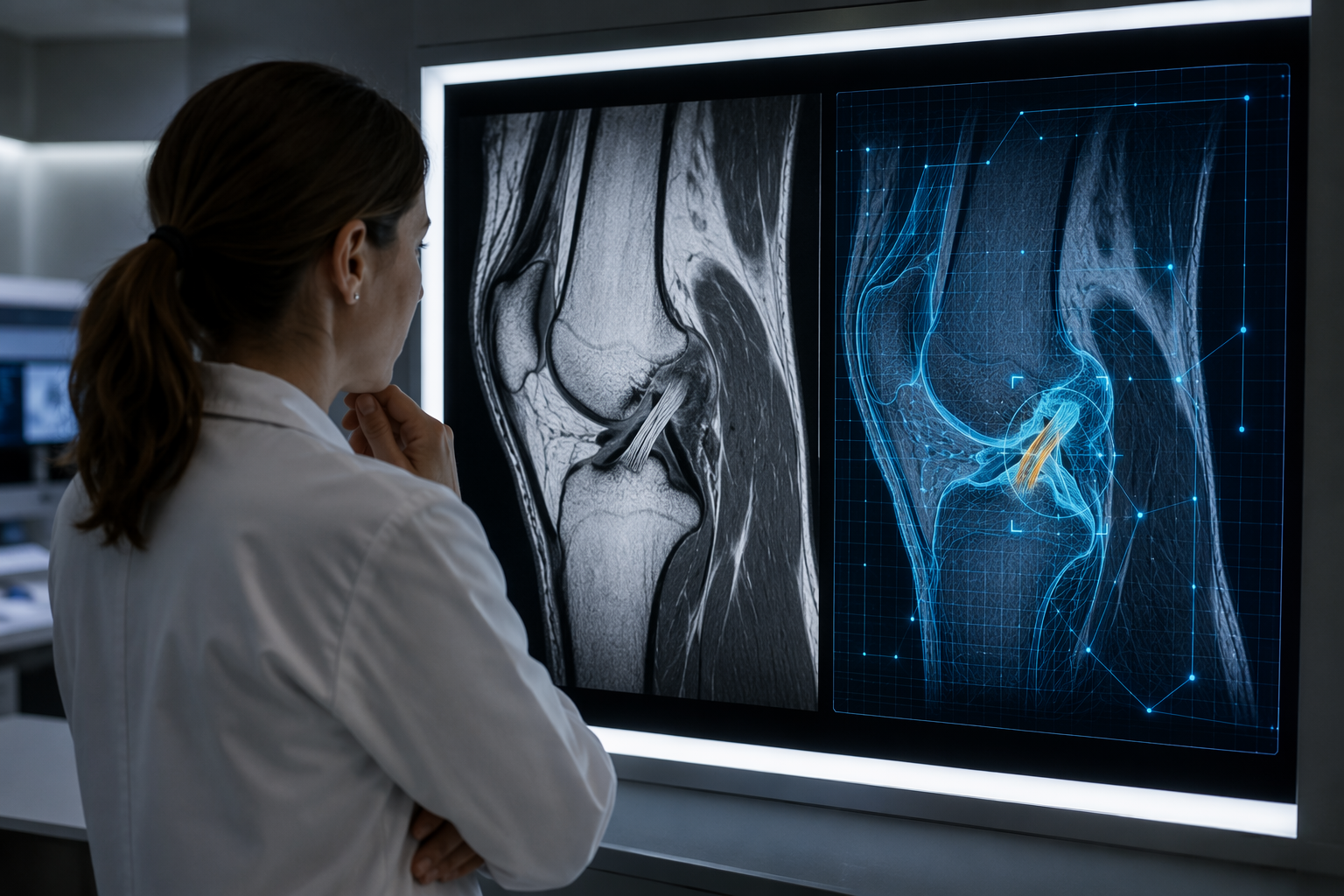
Trusting AI in sports injury assessment is reasonable only when the question is narrow enough to test: Is the ACL torn on this MRI? Is there a fracture on this radiograph? Which implant is shown on this image? For those bounded image-recognition tasks, the evidence is no longer trivial. Deep learning models have reached performance comparable to musculoskeletal radiologists for ACL tear detection on MRI, and other orthopedic imaging models have reported high accuracy for rotator cuff tears, meniscal injuries, fracture detection, and implant identification in defined datasets.[1]
That is different from saying AI imaging tools are ready to run unsupervised across a real clinic. The stronger reading is more cautious: some imaging models can assist clinicians on specific diagnostic or measurement tasks, but the broader evidence base remains uneven, with a regulatory landscape that has moved faster than prospective clinical validation.
The evidence is strongest when the task is specific
The most clinically useful AI studies in sports imaging tend to avoid grand claims. They train a model against a defined imaging task, compare it with human readers or reference labels, and report whether the model finds the abnormality accurately enough to matter. That framing suits musculoskeletal imaging because many sports injuries are image-pattern problems before they become treatment decisions.
ACL tear detection is the cleanest example. MRI already carries much of the diagnostic burden for suspected ligament injury, and an algorithm that flags a complete tear, partial tear, or intact ligament can be tested against expert interpretation. In the evidence summarized in the orthopedic sports medicine literature, deep learning models for ACL tear detection achieved accuracy comparable to musculoskeletal radiologists.[1] That does not make the model a surgeon, and it does not decide whether an athlete needs reconstruction. It does suggest that, for a focused MRI classification problem, AI can reach the level where it may function as a second reader or triage aid.
Rotator cuff and meniscal assessment sit in the same category of plausible assistance. These are common sports and activity-related injuries where MRI interpretation often depends on locating subtle tissue disruption, estimating tear extent, and separating clinically meaningful signal from degenerative or postsurgical change. AI performance in these areas should be judged lesion by lesion and sequence by sequence, not as a blanket statement that an MRI model “understands the shoulder” or “reads the knee.” A model that performs well for supraspinatus tear detection may not be equally dependable for labral pathology, cartilage injury, or postoperative anatomy.
Fracture detection is also a natural fit for computer vision because the immediate clinical question is often binary and time-sensitive: is there a cortical break, avulsion, or stress-related abnormality that should change management? AI tools can help surface abnormalities that are easy to miss when the imaging queue is crowded. Still, the word “fracture” covers a wide range of problems. A displaced clavicle fracture on radiography, an occult scaphoid fracture, and an early stress injury on MRI are not the same diagnostic challenge.
ACLs, cuffs, menisci, fractures: useful signals, not universal proof
The temptation is to add each successful study to a single scoreboard for “AI in sports medicine.” That is a bad habit. Imaging AI earns trust by task, population, scanner or modality, labeling method, and workflow role. A knee MRI model trained to identify ACL tears does not validate an ankle ultrasound tool, a concussion-risk model, or a surgical planning assistant.
| Task | Where AI looks most credible | What still needs checking |
|---|---|---|
| ACL tear detection on MRI | Defined ligament-classification task with performance comparable to musculoskeletal radiologists | External validation, partial-tear handling, scanner variation, and effect on reporting decisions |
| Rotator cuff and meniscal injury assessment | Pattern-recognition support for common soft-tissue injuries | Tear grading, postoperative cases, degenerative overlap, and reader-model disagreement |
| Fracture and stress injury detection | Second-reader or triage support for missed or time-sensitive findings | Performance in subtle, sport-specific overuse injury rather than only obvious fracture datasets |
| Implant identification | Narrow radiographic classification task with very high reported sensitivity and specificity | Whether the task generalizes to less common implants, poor-quality images, and incomplete metadata |
| Anatomic measurement | Reduction of repetitive segmentation and measurement burden | Whether faster measurement changes clinical decisions or outcomes |
Implant identification is an instructive outlier because the reported number is striking and the task is unusually bounded. In radiographic implant identification, one study reported up to 99% sensitivity and specificity.[1] That is impressive. It is also not evidence that orthopedic imaging AI broadly “works.” It shows that when the visual target is constrained, the answer set is finite, and the reference standard is relatively concrete, a model can be extremely good.
That matters in sports medicine because athletes and active patients increasingly arrive with prior surgeries, implants, anchors, and reconstructed anatomy. Correct implant identification can help when operative records are missing or when revision planning depends on knowing what is already in the body. But the clinical consequence is different from diagnosing a new ACL tear. The model is classifying hardware, not weighing pain, instability, rehabilitation goals, and surgical risk.
Measurement AI may be clinically boring in the best possible way
Some of the most credible uses may be the least dramatic. Posterior tibial slope measurement is a good example because the task is tedious, clinically relevant, and relatively well suited to automation. In one reported use case, AI achieved a mean absolute error of about 2 degrees and processed images in about 5 seconds, compared with approximately 180 minutes for manual segmentation.[1]
That kind of time difference is not a marketing flourish if a department is actually doing the measurements. Manual segmentation consumes attention that could be used for interpretation, consultation, or procedural work. A fast measurement tool does not need to replace the radiologist to be useful. It needs to produce a value accurate enough for the intended decision, show its work clearly enough to be corrected, and fit into the reporting environment without creating a second documentation burden.
The same principle applies to other quantitative imaging tasks in sports injury assessment: alignment, version, limb length, cartilage thickness, muscle volume, and postoperative change. The value proposition is strongest when AI removes repetitive measurement work while leaving interpretation and clinical judgment with the treating team.
FDA clearance has grown faster than prospective validation
The regulatory picture is where enthusiasm needs a hard stop. A 2026 analysis of FDA-cleared artificial intelligence medical devices in orthopedic surgery identified 70 orthopedic AI/ML devices cleared as of February 2025.[2] The growth curve was steep: the 3-year moving average of annual clearances rose from 3.0 in 2017-2019 to 16.6 in 2022-2024, a 5.5-fold increase.[2] Since 2022, deep learning accounted for 57.3% of approvals.[2]
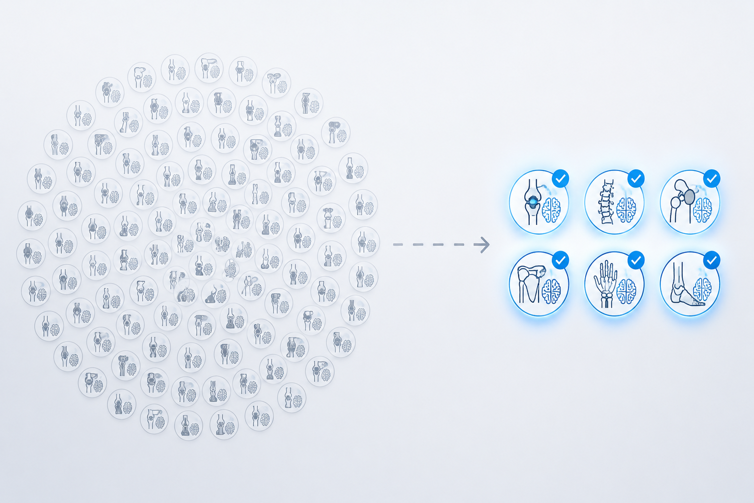
The subspecialty distribution also matters. Spine represented 42.9% of cleared orthopedic AI/ML devices, and hip/knee represented 20.0%.[2] Those categories overlap with sports medicine only in part. A clearance landscape dominated by spine and arthroplasty does not automatically tell a sports medicine clinic how much to trust a model for hamstring injury, cartilage defect characterization, ankle instability, or return-to-play imaging.
The more uncomfortable finding is validation. In the same analysis, only 8.6% of orthopedic AI/ML devices had been validated through prospective clinical trials, while 22.8% lacked any clinical testing.[2] There was improvement over time: the proportion of devices with no clinical testing dropped from 62.2% in 2017-2019 to 19.7% in 2022-2024.[2] That trend is encouraging, but it does not erase the current gap between clearance and real-world evidence.
One reassuring signal from that analysis is safety surveillance: no orthopedic AI/ML device had been subject to a device recall or adverse event report as of the study date, compared with a 10% recall rate for all AI/ML devices and 17.8% for non-AI orthopedic devices.[2] That should be interpreted carefully. Absence of reported recalls is not the same as proof of clinical benefit, and under-recognition or under-reporting of AI-related workflow harm remains possible.
What clearance does not answer
For a clinician deciding whether to rely on a sports imaging AI tool, FDA clearance answers a narrower question than many people assume. A cleared device has passed a regulatory pathway for a specified intended use. It has not necessarily shown that it improves diagnostic accuracy in the local department, shortens time to treatment, reduces missed injuries, improves athlete outcomes, or decreases cost.
That distinction is especially important for 510(k)-style authorization, where substantial equivalence to a predicate device can support market entry without requiring a prospective trial proving better patient outcomes. The regulatory label may be appropriate and still leave the treating team with the practical questions: Who sees the AI output? When does it appear? Can it be ignored? How are disagreements documented? Who is liable if the model misses the subtle finding that the radiologist also overlooks?
The Lee et al. device analysis is valuable because it quantifies this problem rather than merely warning about it.[2] It is still one study. Its conclusions deserve attention, not overextension. A replicated analysis using updated FDA data, device labeling, postmarket surveillance, and published validation studies would make the field easier to judge. As of mid-2026, the best available picture is enough to justify caution: clearance is not a synonym for prospective clinical dependability.
Retrospective accuracy is not the same as workflow performance
Most impressive imaging AI results come from retrospective evaluation. That design is useful and often necessary early in development. It can show whether a model recognizes signal in a labeled dataset. It cannot fully show what happens when the model is dropped into a busy practice with different scanners, protocols, injury prevalence, patient demographics, image quality, and reporting habits.
A model can lose value in several ordinary ways. It may flag too many low-confidence findings and slow the reader. It may perform worse in adolescents, older recreational athletes, postoperative knees, or sports underrepresented in the training data. It may create automation bias, nudging a tired reader away from an abnormality outside the model's intended target. It may produce a heat map that looks persuasive but does not explain enough to resolve a disagreement.
These are not philosophical objections. They are implementation questions. A radiologist signing a report cannot cite a model's retrospective area-under-the-curve as a substitute for explaining why the ligament is torn or intact. An orthopedic surgeon planning treatment needs to know whether the AI output changes confidence, timing, or surgical planning. A sports medicine clinician counseling an athlete needs language for uncertainty, especially when symptoms and imaging do not align.
Stress injury and overuse detection need a lighter touch
Stress injuries are appealing territory for AI because earlier detection could change training, loading, and return-to-sport decisions. The available literature includes work describing AI-driven medical image analysis as promising for earlier detection of stress fractures and other overuse injuries.[3] The caveat is important: one Scientific Reports source discussed a proposed BINN+ASMS framework benchmarked on general-purpose datasets including CamVid, MSRA10K, DUT-OMRON, and NYU Depth V2, rather than being built entirely on direct sports injury clinical imaging data.[3]
That kind of benchmark can be technically informative without being clinically decisive. A sports injury model should ultimately be tested on the imaging problem it claims to solve: the relevant modality, the relevant anatomy, the relevant stage of injury, and the relevant patient mix. For stress injury, that means proving performance in subtle and early cases, not only in image-analysis tasks that are easier to label or more available for training.
Where AI belongs in the clinical chain
The safest near-term role is assistive. AI can pre-read, triage, measure, highlight, compare, or populate structured fields. It can help radiologists find abnormalities sooner and help orthopedic teams receive more consistent quantitative information. It should not silently convert an uncertain image into a falsely certain clinical plan.
- Use AI first for tasks with a defined target, clear reference standard, and published external validation.
- Separate diagnostic support from treatment recommendation; the evidence for image classification is stronger than the evidence for broader clinical decision-making.
- Require local workflow testing before routine reliance, including reader disagreement handling and documentation rules.
- Ask whether the tool was prospectively validated, not only whether it was cleared.
- Monitor misses, false positives, report turnaround, downstream imaging, and clinician behavior after deployment.
The practical adoption question is not whether the model is impressive in isolation. It is whether it changes the work safely. If an ACL tool flags a tear before the radiologist opens the case, does it improve prioritization or merely add another alert? If a fracture detector marks a subtle abnormality, does the reporting radiologist accept, reject, or modify the suggestion? If posterior tibial slope is automatically measured, does the orthopedic surgeon trust the number enough to use it, and can the measurement be corrected when the segmentation is wrong?
Those questions are less glamorous than model architecture, but they are closer to patient care. An AI tool that cannot be integrated into PACS, reporting software, orthopedic planning, or the EHR becomes another screen to check. A tool that shifts liability without clarifying responsibility may be accurate and still unwelcome. A tool that works only in the population on which it was trained may widen errors when used elsewhere.
So, can it be trusted?
Yes, sometimes, and only with the right scope. AI imaging diagnostics can be trusted as assistive tools for defined sports injury assessment tasks when the model has strong validation, a clear intended use, understandable failure modes, and a workflow that keeps clinicians responsible for the final interpretation. ACL tear detection, selected fracture detection, rotator cuff and meniscal assessment, implant identification, and automated measurement are credible areas to watch because the clinical tasks are concrete enough to test.
They should not be trusted merely because they are deep learning tools, cleared devices, or fast. The current evidence supports careful clinical integration, not blanket confidence. Until prospective, multi-center, workflow-aware validation becomes routine, the dependable position is to treat AI as a capable second set of eyes for specific musculoskeletal imaging questions—not as routine clinical infrastructure that has already proven it improves sports injury outcomes.
References
- Artificial Intelligence and its Current Role in Clinical Outcome Prediction, Musculoskeletal Imaging, and Economic and Ethical Considerations within Orthopedics and Sports Medicine, PMC, https://pmc.ncbi.nlm.nih.gov/articles/PMC13076830/
- FDA-Cleared Artificial Intelligence Medical Devices in Orthopaedic Surgery, JAAOS, 2026, https://pmc.ncbi.nlm.nih.gov/articles/PMC12879955/
- AI-driven medical image analysis for sports injury diagnosis, Scientific Reports, https://pmc.ncbi.nlm.nih.gov/articles/PMC12644761/
Comments
Join the discussion with an anonymous comment.