
Legionnaires’ disease enters the hospital through an ordinary door: community-acquired pneumonia with fever, cough, dyspnea, and sometimes gastrointestinal or neurologic features. That is the problem. The patient does not arrive labeled as a waterborne pathogen exposure. The admitting team sees pneumonia, starts down a familiar pathway, and may never order the test that would name Legionella.
That mismatch is why discussions of AI diagnosis tools for Legionnaires’ disease symptoms need some discipline. Symptoms create suspicion; they do not reliably separate Legionella pneumonia from other pneumonias. The diagnostic question is whether newer laboratory and computational tools shorten the distance between nonspecific pneumonia and pathogen identification.
Legionella species are estimated to cause 2–10% of community-acquired pneumonia, yet detected cases remain below that burden; CDC estimates suggest true cases may be 2.7 times higher than reported, and European incidence nearly doubled from 1.4 to 2.2 per 100,000 between 2015 and 2019.[1][2]

Symptoms can raise suspicion, not settle the diagnosis
The clinical pattern is useful only up to a point. Fever, cough, shortness of breath, radiographic pneumonia, diarrhea, confusion, hyponatremia, exposure to building water systems, travel, or an outbreak context may push Legionella higher on the differential. None of that gives a clinician species-level confirmation at the bedside.
This is where diagnostic pathways matter more than diagnostic hunches. If the order set stops at a narrow urine antigen test, the hospital has already accepted a blind spot. If the pathway includes broader molecular testing when Legionella is plausible, the organism has a better chance of being found early enough to affect antibiotics and infection prevention work.
The consequence is not theoretical. In the cited review, delayed appropriate antibiotics beyond 24 hours after admission were associated with ICU admission in 80.4% of cases, compared with 54.2% when appropriate treatment was timely.[1]
The urine antigen test explains much of the missed-case problem
The urine antigen test is popular for understandable reasons. It is fast, familiar, noninvasive, and operationally simple. In European diagnoses, it is used in roughly 90% of cases.[1] For a busy pneumonia admission, those virtues are real.
But the test is narrow. Standard UAT detects L. pneumophila serogroup 1, with reported sensitivity around 75–86%, and misses other serogroups and Legionella species.[1][2] A negative result therefore does not mean the patient does not have Legionnaires’ disease. It may mean the hospital looked only where the light was easiest.
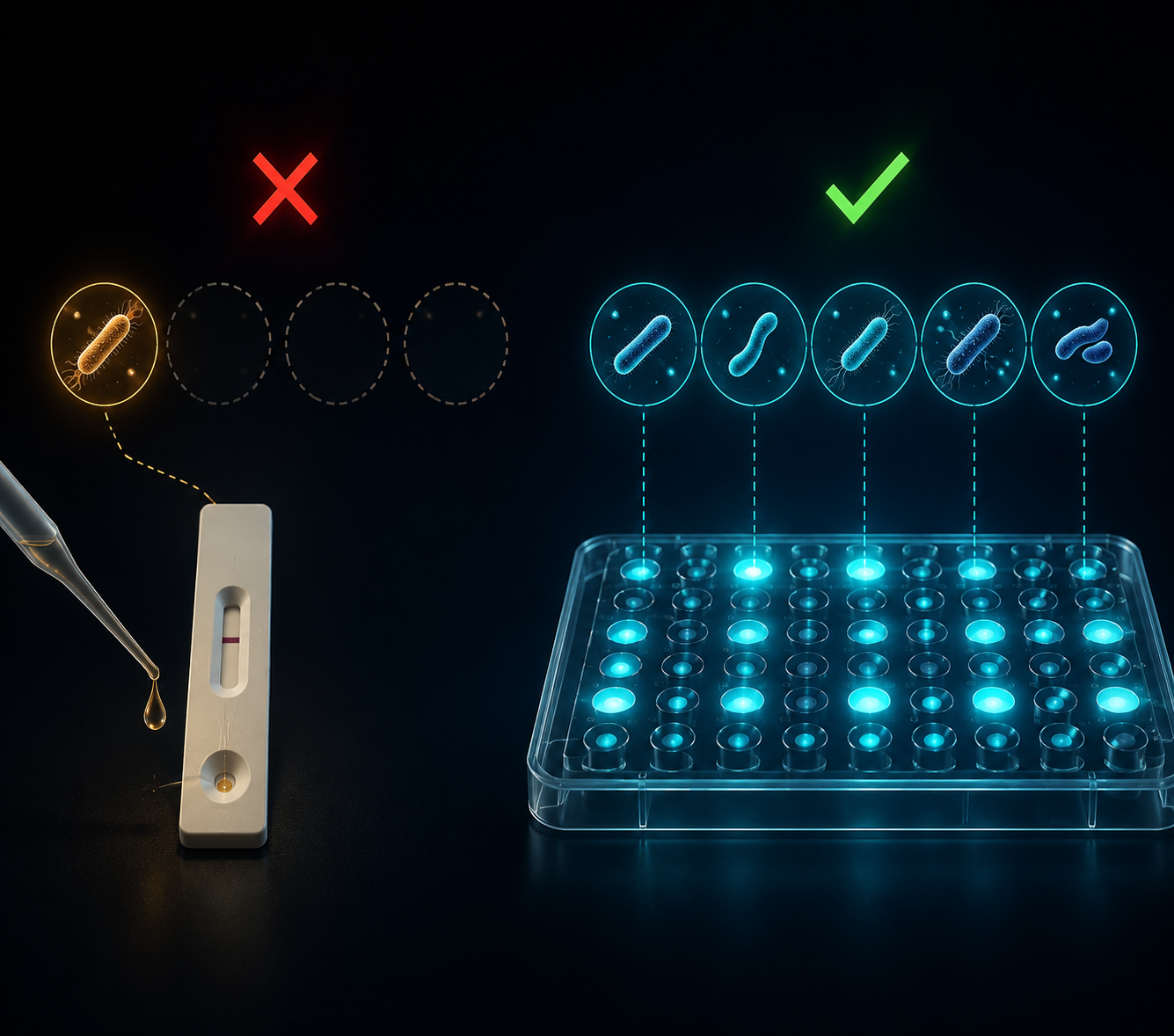
The clinical irritation here is not that UAT exists. It is that a narrow test can become the de facto definition of the disease. If detection depends mainly on serogroup 1 antigen in urine, then the reported epidemiology will tilt toward what that test can see. Cases due to other species or serogroups are easier to miss, especially when symptoms offer no reliable rescue.
| Diagnostic tool | What it detects | Reported role in European diagnoses | Main limitation |
|---|---|---|---|
| Urine antigen test | Primarily L. pneumophila serogroup 1 | Roughly 90% | Misses non-serogroup 1 and other Legionella species; sensitivity about 75–86% |
| Multiplex PCR | All known Legionella species | Less than 10% | Requires appropriate respiratory sampling and adoption into ordering pathways |
Multiplex PCR is the most practical improvement in detection
Multiplex PCR changes the diagnostic question from “does this urine sample show antigen from the most commonly detected serogroup?” to “is Legionella genetic material present, including species the urine antigen test will not detect?” That is not AI magic. It is better coverage, and in pneumonia diagnostics, better coverage is often the difference between a named pathogen and another culture-negative admission.
The reported performance is clinically meaningful: PCR achieves about 90% sensitivity and can detect all known Legionella species, yet it accounts for less than 10% of European diagnoses.[1] That gap is hard to defend if the goal is to reduce missed cases rather than simply process familiar tests.
The specimen issue matters. PCR depends on respiratory material, and getting that specimen is not always as automatic as collecting urine. Patients may not produce sputum, lower respiratory sampling may not be justified in every admission, and local laboratories vary in which panels they run. Those are workflow barriers, not reasons to pretend the narrower test is adequate.
A more sensible pathway is selective broadening. A patient with community-acquired pneumonia and features that keep Legionella on the differential should not be closed out by a negative UAT when the clinical stakes are high or the epidemiologic context is suspicious. PCR is most useful when it is placed where decisions happen: early enough to influence macrolide or fluoroquinolone coverage, and visible enough to alert infection prevention if multiple cases suggest a shared exposure.
This is also where “AI-enhanced” language can obscure more than it helps. The immediate gain is not a black-box model reading a patient’s symptom list. It is a diagnostic system that pairs clinical suspicion with broader pathogen detection and routes the result back to clinicians before treatment decisions have hardened.
Where machine learning becomes more convincing: outbreak attribution
Genomics-based machine learning belongs in a different part of the pathway. It is not the first tool I would expect to diagnose the febrile patient in the emergency department. Its stronger role begins after isolates or genomes are available, when the question becomes whether clinical cases connect to each other or to an environmental source.
One Legionella pneumophila source-attribution study used a pan-genome SNP approach with 479,480 SNPs, compared with 221,214 core SNPs, and evaluated 20 outbreak groups from Australia, the United Kingdom, and the United States.[3] The machine learning classifier achieved a mean F1 score of 0.74, outperforming standard phylogenomic analysis at 0.50 and cgMLST at 0.56; it reached perfect F1 in 13 of the 20 outbreak groups.[3]

The useful point is the pan-genome signal. Core-genome methods restrict attention to regions shared across isolates. A pan-genome approach can use a wider set of variation, which may help when source attribution depends on distinctions that standard comparisons flatten. The performance comparison matters because outbreak teams do not need elegant genomics in the abstract; they need to know whether a patient isolate plausibly points to a cooling tower, plumbing system, spa, or other environmental source.
The boundary is just as important. The study’s training geography was limited to three countries, and performance in other regions or across broader sequence diversity still needs validation.[3] A classifier that improves attribution in one evidence base is not automatically a universal Legionella source oracle. It is, however, a serious public health tool when deployed with genome generation, environmental sampling, and epidemiologic investigation.
This is where related work on AI for bacterial detection in hospital water systems fits naturally: clinical diagnosis and environmental surveillance are separate workflows, but Legionella control depends on both. A hospital can identify a case quickly and still fail the next patient if the source investigation stalls.
Imaging AI is not ready to distinguish Legionella at the species level
Chest imaging AI has a plausible future in pneumonia triage and pattern recognition, but that is not the same as Legionella diagnosis. The current evidence base described here does not support treating AI-assisted chest imaging as validated for species-level differentiation of Legionella pneumonia. Most such work remains closer to broad pneumonia detection than pathogen-specific identification.
That distinction matters at the bedside. A radiology model that flags pneumonia may speed recognition of lung infection, but it does not tell the clinician whether a negative urine antigen test missed a non-serogroup 1 Legionella infection. It also does not replace respiratory sampling, molecular testing, culture when available, or public health linkage during clusters.
The practical line is straightforward: multiplex PCR is the evidence-ready improvement for clinical detection; genomics-based machine learning is performance-relevant for outbreak attribution once genomes exist; imaging AI should remain a research tool for Legionella-specific differentiation until validation catches up.
References
- Legionnaires’ Disease: Update on Diagnosis and Treatment. PMC.
- Clinical Features of Legionnaires’ Disease and Pontiac Fever. Centers for Disease Control and Prevention.
- High performance Legionella pneumophila source attribution using genomics-based machine learning classification. Applied and Environmental Microbiology.
Comments
Join the discussion with an anonymous comment.