
The celebrity version of AI preventive care is easy to caricature: polished clinics, high-income patients, a scan marketed as reassurance. That caricature misses the more interesting clinical problem. Some of these services are built around serious imaging infrastructure, high-throughput acquisition, and machine-learning workflows that look familiar to anyone watching radiology AI mature. At the same time, the American College of Radiology does not recommend routine total-body screening MRI for average-risk asymptomatic adults, largely because incidental findings and false positives can create downstream work without proven outcome benefit.
That is the tension worth examining. The question is not whether famous investors or patients make a scan clinically meaningful. It is what kind of evidence would have to connect detection, follow-up, treatment, and patient outcomes before AI-enabled whole-body screening becomes more than an impressive concierge service.
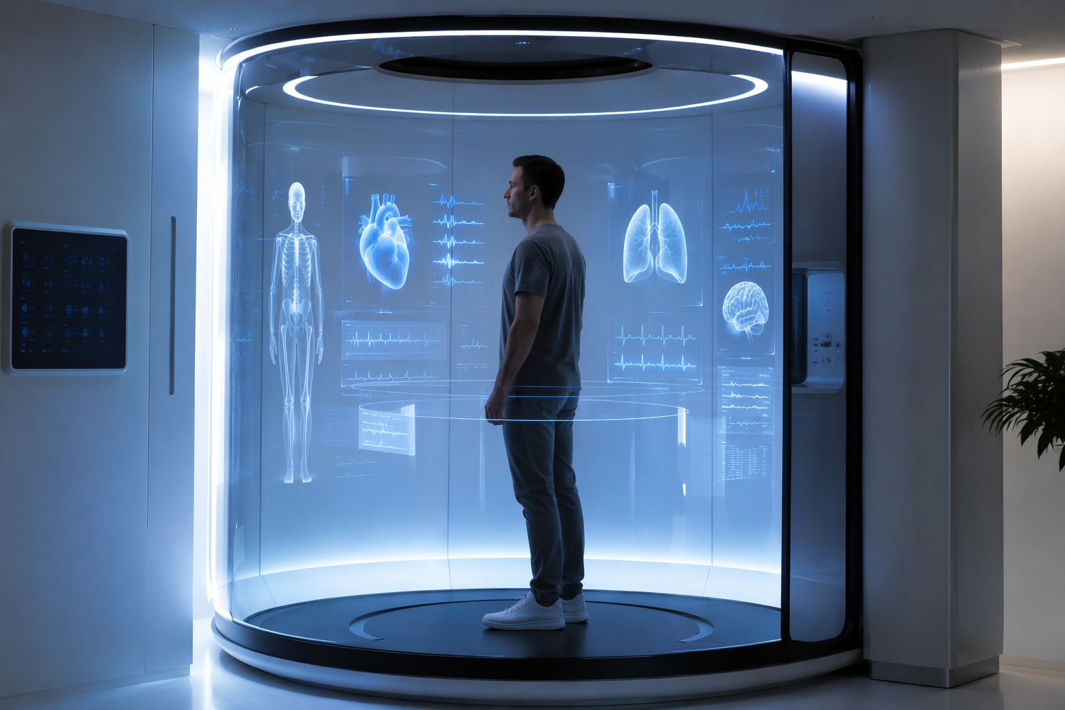
The phrase celebrity family health monitoring slightly overstates what these clinics usually offer. Neko Health and Prenuvo are primarily individual screening services for asymptomatic adults, not family monitoring systems in the sense used for consumer wearables or home health dashboards. The family angle matters mostly because celebrity adoption can normalize a pattern of repeated preventive scanning across affluent households. The clinical evidence still has to be judged scan by scan, finding by finding, and pathway by pathway.
Three Claims That Should Not Be Blended Together
The confusion starts when three related claims are treated as one. First, AI-assisted imaging is clinically real. Second, whole-body or multi-system screening can find abnormalities in people who feel well. Third, finding those abnormalities improves outcomes enough to justify routine screening in average-risk adults.
Only the first claim is already broadly established. FDA-cleared imaging AI is no longer a speculative category: ClinicalMind’s analysis of FDA AI device clearances in 2025 reported 295 AI-enabled medical devices cleared in 2025, 1,451 cumulative authorizations through 2025, and radiology or imaging as the dominant category. That clearance context matters because it means image-analysis algorithms, triage tools, reconstruction techniques, and measurement systems are entering regulated clinical workflows at scale.
But clearance is not a transferable blessing. A device can be cleared for a specific imaging task, population, modality, workflow, or performance claim. That does not prove that a concierge screening program using multiple technologies improves mortality, reduces morbidity, or produces acceptable downstream harm in average-risk asymptomatic adults. Intended use is not a paperwork detail; it is the boundary around what has actually been evaluated.
The second claim is also plausible, and in some reports more than plausible. Whole-body imaging and multi-sensor exams will find lesions, vascular markers, masses, skin findings, metabolic signals, or other abnormalities that a routine visit would miss. The harder question is what happens next: how many findings are actionable, how many are false positives, how many trigger surveillance or biopsy, and how many lead to interventions that change outcomes rather than simply move diagnosis earlier.
What Neko Health and Prenuvo Are Actually Counting
Neko Health and Prenuvo are useful examples because they are not the same technical product. Neko’s model is described as a rapid multi-sensor scan rather than a whole-body MRI. Reporting on the clinics describes a 10-minute scan using more than 70 sensors, generating 50 million data points and building an AI-enabled “digital twin” intended for longitudinal comparison. In a Stockholm cohort of 2,707 patients, the same report says 14.1% needed previously unknown medical treatment and 1% received interventions described as life-saving for severe conditions.[1]
Prenuvo sits closer to the radiology controversy because it is built around full-body MRI. The reported operational scale is substantial: more than 170,000 scans across 26 clinics, approximately 1.3 billion data points per full-body MRI scan, about $100 million in annual revenue, and a company-reported life-saving detection rate around 5%.[1]
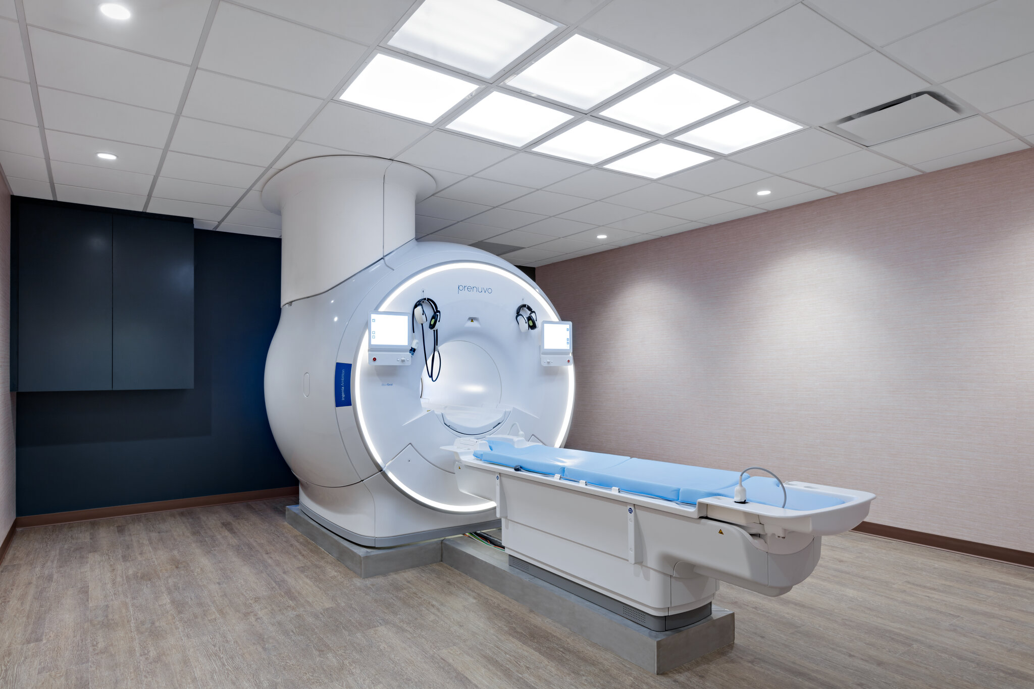
Those numbers are too large to wave away. A 14.1% treatment yield in an asymptomatic screening population, if independently reproduced with clear adjudication, would deserve serious clinical attention. So would a true 5% life-saving detection rate. The caution is that these are not equivalent to prospective evidence that routine screening improves outcomes. They are operational signals, and much of what is publicly available comes through company-reported or non-peer-reviewed channels.
The wording also matters. “Needed treatment” is not the same endpoint as reduced mortality. “Life-saving” is a high-value claim, but it requires a counterfactual: what would likely have happened without the scan, and over what time horizon? A malignant tumor found at an early stage, an aneurysm identified before rupture, and a benign lesion sent for repeat imaging all sit in very different clinical categories. A screening program can report all of them as detections, but clinicians need the denominator and the disposition of each branch.
| Claim Type | What It Can Support | What It Cannot Prove Alone |
|---|---|---|
| FDA-cleared imaging AI | A specific device or algorithm met regulatory requirements for its cleared intended use. | A whole screening program improves outcomes in average-risk asymptomatic adults. |
| High detection yield | The clinic found previously unknown abnormalities or treatment needs in screened patients. | The benefits exceed false positives, overdiagnosis, anxiety, cost, and downstream procedures. |
| Longitudinal digital twin or repeat-scan model | The system can compare a person’s current measurements with prior measurements over time. | Earlier detection changes hard outcomes without unnecessary intervention. |
| Professional society caution | Existing evidence is not sufficient for routine use in the specified population. | The technology can never become useful under better-defined risk or trial conditions. |
Why Longitudinal AI Comparison Is More Than a Marketing Detail
The most clinically interesting part of the Neko-style model is not the sleek scan itself. It is the idea that repeated exams could turn a single cross-sectional screening event into a longitudinal measurement system. A one-time abnormal value forces a clinician to decide whether it is disease, noise, normal variation, or a benign outlier. A repeated measurement can show whether the signal is stable, resolving, or changing in a pattern that deserves escalation.
That is where AI may have a legitimate role. Computer vision systems can segment structures, quantify change, standardize measurements, compare current and prior images, and flag patterns that may be difficult to track manually at scale. For readers who want the imaging mechanics rather than the clinic narrative, ClinicalMind’s explainer on computer vision AI in imaging covers the underlying workflow more directly.
Longitudinal comparison also cuts both ways. It can reduce uncertainty when a finding remains unchanged, but it can also create a surveillance obligation. Once a person has a small indeterminate lesion, a borderline vascular measurement, or a signal that is “probably benign,” someone has to decide when to re-image, when to refer, and when to stop. The clinic can generate the first report quickly; the health system absorbs the ambiguous tail.
The ACR Objection Is About the Screening Strategy, Not About AI Itself
The ACR position against routine total-body screening MRI for average-risk asymptomatic adults is sometimes read as a rejection of innovation. That is too blunt. The objection is more procedural: if a screening test is offered broadly to people without symptoms or defined risk, the evidence burden is higher because most people screened will not have the target condition.
In that setting, even a technically excellent scan can create harm. Incidental findings can lead to repeat imaging, specialist visits, biopsy, surgery, anxiety, and cost. False positives are not merely statistical artifacts; they become phone calls, referrals, and clinical responsibility. The radiologist or primary care clinician downstream of the luxury scan has to explain probability after the marketing encounter has already framed detection as prevention.
This does not mean the ACR position should freeze the field. Guidelines follow evidence; they do not generate it. But they do identify the current burden of proof. A clinic reporting high detection yield has not yet answered the guideline concern unless it also reports adjudicated outcomes, false-positive burden, follow-up completion, complication rates, and comparison against usual care or risk-targeted screening.
What a Credible Trial Would Need to Show
A prospective evaluation would not have to pretend that whole-body AI screening is a single intervention. It would need to specify the population, the scan protocol, the AI components, the reporting thresholds, and the follow-up pathway before screening begins. Otherwise, a positive-looking result can be assembled after the fact from whatever findings are most persuasive.
The primary endpoint is the hard part. Mortality may be too slow or underpowered for early trials, but a study still needs clinically meaningful outcomes rather than only counts of abnormalities. Stage shift for selected cancers, urgent interventions for validated high-risk findings, avoided acute events, quality-of-life effects, downstream procedure rates, and cost per actionable diagnosis would all be more informative than a global detection percentage.
The comparator matters just as much. Whole-body MRI or multi-sensor screening should be tested against something: usual preventive care, guideline-based screening, risk-stratified imaging, or a narrower targeted protocol. Without a comparator, the evidence can show that the clinic found things. It cannot show whether the same people would have done better, worse, or no differently under existing care.
- Population: average-risk asymptomatic adults should be analyzed separately from people with genetic risk, prior cancer, concerning symptoms, or strong family history.
- Finding categories: malignant, premalignant, vascular, inflammatory, benign incidental, indeterminate, and false-positive findings should not be collapsed into one success metric.
- Follow-up: the study should track who receives additional imaging, procedures, specialist visits, treatment, surveillance, or no action.
- Harms: anxiety, complications, unnecessary procedures, radiation from follow-up CT, and financial burden should be counted, not treated as secondary inconvenience.
- Validation: claims about life-saving detection should be independently adjudicated rather than inferred from the seriousness of the diagnosis alone.
Where the Celebrity Clinic Model Adds Real Friction
Concierge preventive screening changes the clinical workflow before the evidence question is settled. Patients arrive with a report, a list of findings, and often an expectation that the scan has revealed something medicine would otherwise have missed. The receiving clinician inherits the task of translating a broad screening result into a risk-specific plan.
There is also an access issue, though it is not the central evidentiary problem. If a service is expensive, concentrated in major cities, and marketed through celebrity proximity, the early user base will not represent the general screening population. That limits what can be inferred from operational yield. Detection rates in self-selected, affluent, health-conscious patients may not generalize to broader primary care populations.
Still, dismissing the model as vanity medicine is too easy. A high-throughput clinic that repeatedly captures structured imaging and physiologic data could become a valuable evidence engine if it commits to independent validation. The same operational scale that makes the marketing powerful could also make the science better, provided the data include negative findings, downstream harms, and long-term outcomes rather than only compelling detections.
What Is Supported Now
The supported claim is narrow but meaningful: AI-enabled imaging and measurement technologies are mature enough to be evaluated seriously in preventive screening workflows. The broader AI-in-care landscape is already diverse, as shown in ClinicalMind’s review of AI in healthcare by specialty. Radiology is one of the most developed clinical areas because imaging produces structured digital inputs and measurable outputs.
The supported clinic-level claim is also narrower than the marketing sometimes implies: these programs report non-trivial detection yields in screened asymptomatic people. Neko’s reported Stockholm figures and Prenuvo’s reported operational scale are not trivial signals.[1] They justify evaluation, publication, and scrutiny.
What remains unsupported is the leap from detection to routine screening recommendation for average-risk asymptomatic adults. That leap requires prospective evidence that the benefits of earlier detection outweigh false positives, incidental findings, overdiagnosis, downstream procedures, and cost. Until that evidence exists, FDA clearance for component technologies and company-reported yield should be kept in their proper lanes.
The unresolved question is therefore not whether the scans are technologically impressive. Many are. It is whether the current evidence justifies routine use outside carefully defined risk groups, follow-up protocols, and independent validation. On that question, the controversy remains open for good reasons.
Comments
Join the discussion with an anonymous comment.