
Intervertebral disc disease in chondrodystrophic dogs is hard to screen because the relevant signals do not mean the same thing. A genetic result can identify inherited risk from birth, a lateral radiograph can suggest narrowed disc spaces, and MRI can show spinal cord compression, but those are not interchangeable findings. In that sense, AI in veterinary medicine for genetic spinal disorders is most useful when it helps sort risk from current disease instead of collapsing both into one label.
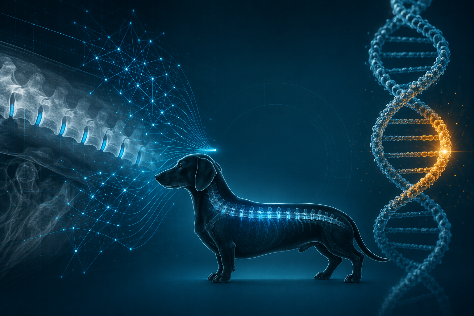
What the radiograph model actually measures
In Park et al.’s single-center study, a 1D-CNN was trained on 241 X-rays from 142 dogs to detect narrowed intervertebral disc space sites on caudal thoracic and lumbar lateral radiographs. The model reached an AUC of 0.837, with 81.5% sensitivity and 95.6% specificity, and its agreement with veterinary clinicians was substantial at kappa = 0.780. It processed each image in 0.104 seconds, compared with 12.2 seconds for clinicians, which makes it about 117 times faster on this task [1].
| Signal | What it measures | What it can support | What it cannot decide |
|---|---|---|---|
| AI radiograph | Narrowed disc space sites on lateral X-rays [1] | A suspicion tier that can prioritize closer review or advanced imaging | Whether the spinal cord is compressed today |
| FGF4 retrogene test | Inherited CFA12 retrogene risk linked to chondrodystrophy and IVDD [2][3] | Lifelong risk-aware monitoring in predisposed dogs | When clinical disease will begin or how severe it will be |
| MRI | Direct assessment of spinal cord compression | Confirmation of the anatomical problem when signs or screening raise concern | Inherited predisposition by itself |
That distinction matters because the model is still reading a radiographic proxy, not the cord itself. The same study reported only moderate agreement, kappa = 0.468, with MRI-confirmed spinal cord compression, which is exactly the kind of result that keeps the word diagnosis out of the AI section. The screen is useful when it identifies dogs that deserve a better next step; it is not useful if it is treated as the next step itself [1].
Why the genetics signal carries different weight
The FGF4 retrogene on CFA12 is not just a background association. In the genetics literature summarized by Dickinson and Bannasch, it is associated with a very large odds ratio for Hansen Type I IVDD, 51.23 with a 95% CI of 46.69–56.20 [2]. Breed-level allele frequencies are also high: Beagles, Cavalier King Charles Spaniels, Dachshunds, and French Bulldogs are all reported above 0.90 in the UC Davis summary of the field [3].
That same UC Davis summary reports that retrogene carriers present for decompressive surgery at a younger mean age, 6.1 years versus 8.5 years in non-carriers, and that histopathological disc degeneration is detectable by 10 weeks of age in homozygous dogs [3]. Those findings support strong biological risk, but they still do not tell a clinician whether a particular dog has clinically meaningful cord compression today. The genetic result says the dog was born into a high-risk category; it does not say when the category becomes disease.
Why the two signals belong in the same screening conversation
The broader veterinary AI literature describes a field that is clearly emerging, but still uneven in validation and clinical deployment [4]. That is the right frame for spinal imaging too. No FDA-cleared or CE-marked veterinary spinal imaging AI is available here as routine clinical software, so the current question is not whether the model is ready to replace radiology; it is whether it can make screening more orderly while the clinician remains responsible for interpretation.
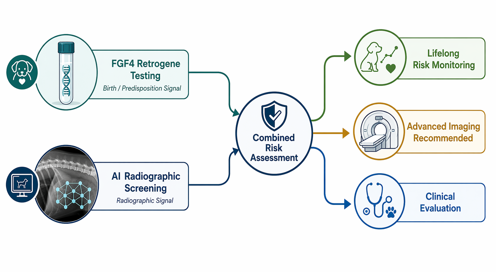
A workable combined workflow is narrow rather than grand. FGF4 testing can identify dogs that need lifelong risk-aware monitoring. AI-assisted radiographic screening can sort lateral spinal films by suspicion and point the concerning ones toward closer clinical review or MRI. The output should be a triage decision, not a disease label.
- If the dog is from a predisposed breed, the genetic test carries the long-horizon risk information.
- If lateral thoracolumbar radiographs are already being taken, the AI model can help standardize which films deserve more attention [1].
- If the AI result is positive, the next question is whether MRI or a neurologic workup changes management, not whether the dog already has definitive disease [1].
No published study has yet tested AI radiographic screening and FGF4 testing as a single clinical program, so the combined paradigm remains a proposal rather than a validated protocol.
References
- Development of a deep learning model for automatic detection of narrowed intervertebral disc space sites in caudal thoracic and lumbar lateral X-ray images of dogs — Frontiers in Veterinary Science, 2024
- Current Understanding of the Genetics of Intervertebral Disc Degeneration — PMC, 2020
- Unraveling the Genetics of Disc Disease in Dogs — UC Davis
- Applications and Considerations of Artificial Intelligence in Veterinary Sciences — PMC, 2024
Comments
Join the discussion with an anonymous comment.