
Computer vision AI in medical imaging is no longer a speculative category waiting for clinical contact. It is already present in regulated imaging workflows, unevenly distributed across modalities, and numerous enough that the first useful question is not whether it exists, but where it has actually been authorized and what kind of evidence is attached to it.
As of October 2024, a peer-reviewed review of the European and U.S. radiology AI landscape counted 222 commercial AI-based radiology products in the European market, with 213 certified, and more than 700 FDA-cleared AI healthcare algorithms in the United States. Radiology accounted for 76% of those FDA-cleared algorithms, or 527 radiology products in that count.[1]
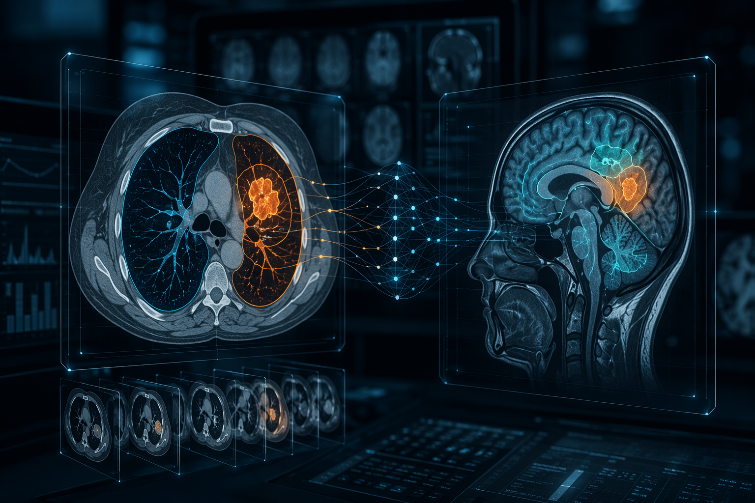
Those figures are snapshot data, not a live inventory. New products may have entered or left the market since late 2024, and FDA device counts vary by inclusion rules. Some industry landscapes report higher totals, including counts above 950, but the conservative peer-reviewed figure is the safer anchor when discussing clinical state rather than vendor momentum.[1][2]
The pattern matters more than the headline number. The market did not simply expand in every direction. In the EU product analysis, first market entries peaked in 2020 with 50 products, while FDA clearance activity peaked in 2023 with more than 80 cleared products; both measures declined afterward, which the review interprets as a move toward maturation rather than disappearance.[1]
Where Imaging AI Has Actually Concentrated
The strongest footprint for computer vision AI in medical imaging is not evenly spread across the reading room. It sits disproportionately in CT and MRI, with neuroimaging and chest imaging leading by subspecialty. That distribution is clinically intuitive: these are high-volume, high-acuity, highly digitized domains where detection, segmentation, quantification, and triage can be framed as device functions.
| Category | Largest concentrations reported as of October 2024 |
|---|---|
| Modality | CT: 89 products; MRI: 66; X-ray: 46; mammography: 16; ultrasound: 10.[1] |
| Subspecialty | Neuroimaging: 73 products; chest imaging: 71; with additional products in musculoskeletal, abdominal, cardiac, and breast imaging.[1] |
| Disease target | Lung cancer: 28 products; stroke: 24; breast cancer: 19.[1] |
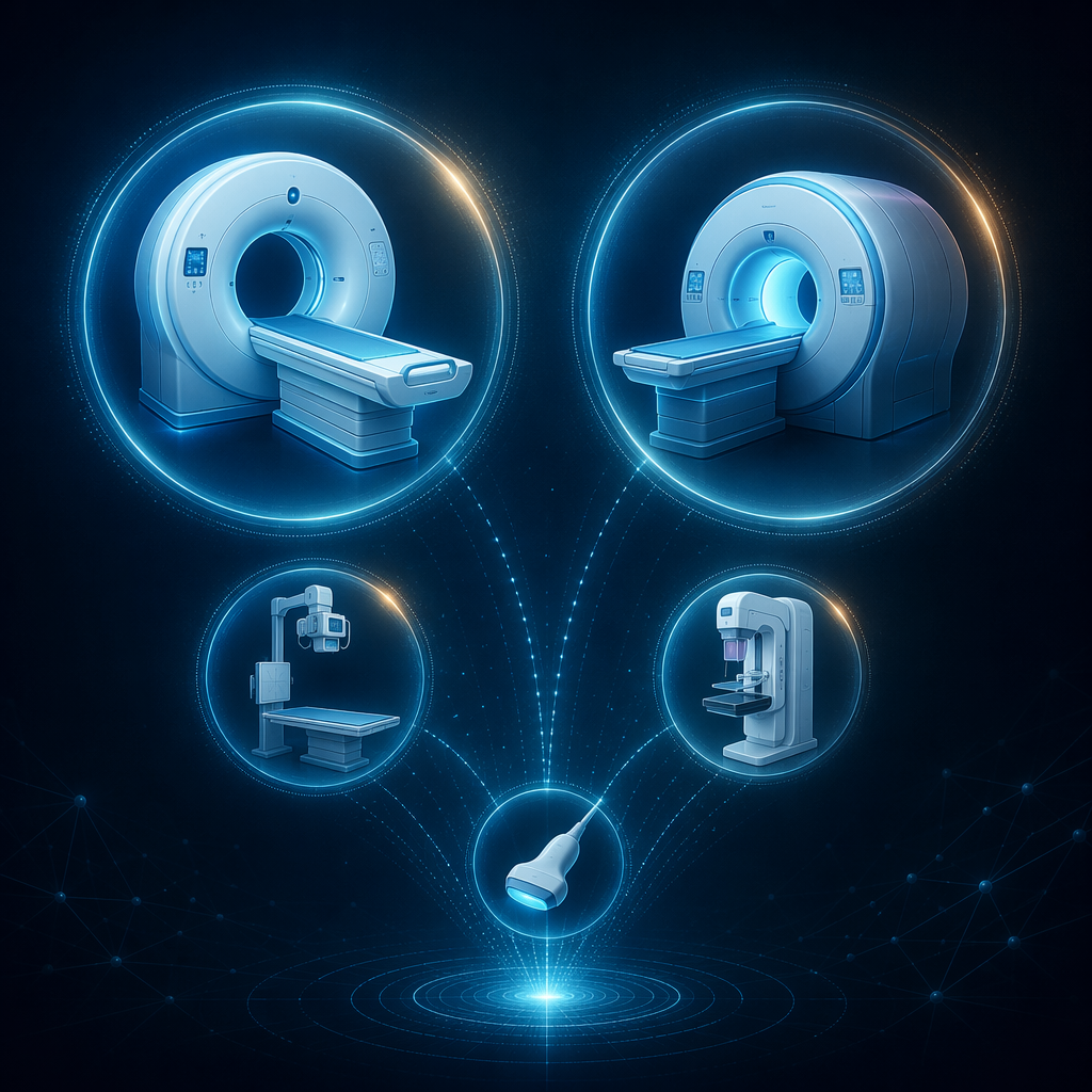
CT’s lead is not surprising. CT produces standardized volumetric data at scale, supports time-sensitive decisions, and lends itself to tasks such as hemorrhage detection, pulmonary embolism flagging, lung nodule identification, fracture detection, and quantitative measurements. A tool that only adds a probability score to a report may be easy to ignore; a tool that changes the order of a worklist, pre-populates a measurement, or sends a stroke alert to a defined clinical team creates a more concrete operational claim.
MRI’s position is different. The modality is rich, multiparametric, and often more variable across protocols. That makes it attractive for segmentation and classification, especially in neuroimaging and prostate imaging, but it also raises the bar for external validation. A model that performs well on one scanner mix, acquisition protocol, or disease-enriched dataset may not behave the same way in a general service list.
Neuroimaging and chest imaging dominate because they combine volume with clinical consequence. Stroke triage is an obvious example: speed matters, the imaging finding has downstream urgency, and the AI output often aims to move a case up the queue. Chest imaging has a different center of gravity, with lung cancer and nodule workflows showing how computer vision can support follow-up, detection, and measurement rather than a single dramatic alert.
Mammography, ultrasound, cardiac imaging, pathology, and prostate imaging matter, but they do not occupy the same market shape in the 2024 radiology product count. Mammography has a narrower product footprint than CT or MRI in that analysis. Ultrasound has fewer counted products, partly because real-world acquisition variability and operator dependence make the modeling and workflow problem harder. Cardiac imaging and pathology extend the same computer vision logic beyond conventional radiology, but their evidence bases and deployment pathways need to be read on their own terms rather than treated as interchangeable with CT triage software.
What These Systems Usually Do
Most clinicians do not need a model-architecture lecture to judge an imaging AI claim. They do need to know what kind of output is being inserted into care. A detection tool flags a suspected finding. A segmentation tool outlines anatomy or pathology. A triage tool changes priority or notification flow. A classification tool assigns a category, probability, or risk group. A measurement tool calculates volume, diameter, density, or change over time.
Those distinctions are not cosmetic. They determine who must act. A segmentation error may slow a radiologist or distort a quantitative biomarker. A false-positive triage alert may interrupt an on-call team. A false-negative triage output may leave a genuinely urgent case in the usual queue. A classification score may shape a biopsy discussion or multidisciplinary review only if the institution has decided how much weight to give it.
Convolutional neural networks remain the familiar backbone for many image-analysis systems, with architectures such as ResNet associated with classification and U-Net associated with segmentation. Newer visual AI discussions increasingly include vision transformers and foundation-model-style approaches, but architecture alone says little about clinical readiness unless it is paired with validation, workflow testing, and a clear intended use.[2]
Clearance Is Meaningful, but It Is Not Clinical Proof
Regulatory clearance is not trivial. It means a product has crossed a defined medical-device threshold for a specified intended use. For hospitals, that matters: clearance constrains labeling, procurement language, risk classification, and governance. It is one reason a cleared worklist triage tool belongs in a different conversation from a promising research model posted with an impressive receiver operating characteristic curve.
But clearance should not be mistaken for proof that a tool improves patient outcomes, reporting quality, turnaround time, radiologist workload, or downstream management in a particular institution. The same 2025 review that counted the regulated product landscape also emphasized that most products still lack rigorous prospective and multi-center clinical trials, and that many evaluations rely on limited study designs.[1]

This is where many product discussions become too loose. A retrospective test set can show that a model detects a finding under controlled conditions. It does not necessarily show that the model improves care when integrated into PACS, routed through an alerting layer, reviewed by radiologists with different experience levels, and used on patients whose scanners, protocols, comorbidities, and prevalence differ from the development data.
The evidence gap is not a reason to dismiss the field. It is a reason to separate questions that are often collapsed into one sales sentence: Is the software authorized? Does it perform on an external dataset? Does it perform across sites? Does it change clinician behavior? Does that behavior improve a clinical or operational endpoint? Does it create new work somewhere else in the system?
The Ground Truth Problem
Computer vision papers often look more definitive than they are because the reference standard is treated as settled. In imaging, ground truth may be a single radiologist annotation, a consensus read, pathology, follow-up imaging, clinical adjudication, or some combination of these. Those choices change what the model is being judged against.
The radiology AI product review specifically criticized reliance on single-annotator ground truth and incomplete statistical reporting, including missing confidence intervals in evaluations.[1] That combination should make any quality committee pause. A point estimate without uncertainty can look clean in a slide deck and still be too brittle for comparison across products, patient groups, or sites.
For a detection model, a single annotator may miss subtle disease or mark borderline findings differently from local practice. For a segmentation model, two experts may disagree at the boundary of tumor, edema, hemorrhage, plaque, or organ capsule. For a triage model, the technically correct label may still be clinically incomplete if the system does not define who receives the alert, how quickly they see it, and what happens when the alert is wrong.
Confidence intervals, external validation, and population reporting are not academic decoration. They are how a reader sees whether an accuracy claim is stable, whether performance was tested outside the development environment, and whether important subgroups were visible rather than averaged away.
Bias Is a Deployment Issue, Not Just a Dataset Issue
Bias in imaging AI is often described as a training-data problem, and it is that. But once software reaches clinical workflow, bias also becomes a routing, review, monitoring, and accountability problem. If performance differs by scanner, site, body habitus, sex, race, age, disease prevalence, or acquisition protocol, the consequence is absorbed by patients and clinicians rather than by the validation spreadsheet.
Reviews of machine learning bias in medical imaging have emphasized that biased data, label quality, underrepresented populations, and inadequate external validation can all affect model behavior.[3] A separate 2025 review focused on radiology AI bias describes detection, avoidance, and mitigation as continuing needs rather than solved implementation details.[4]
The practical implication is straightforward: a site should not ask only whether an AI tool was accurate in a published dataset. It should ask whether the evaluated population resembles its own, whether subgroup performance was reported, whether failure modes are described, and whether local monitoring can detect drift after deployment.
A Stronger Evidence Example: Prostate MRI
The prostate MRI literature gives a useful example of what more serious evaluation can look like. The PI-CAI study, published in The Lancet Oncology in 2024, prospectively evaluated AI for clinically significant prostate cancer detection on MRI and compared AI performance with radiologists.[5]
That does not mean every prostate AI system is clinically interchangeable with the evaluated system, or that the findings automatically generalize to all acquisition protocols and populations. It does show why prospective design and reader comparison change the quality of the conversation. The question moves from whether a model can score images to whether it can stand up against a clinically relevant comparator in a defined diagnostic task.
That kind of study is still not the norm across the broader commercial imaging AI landscape. It is better treated as a benchmark for the kind of evidence clinicians should want, not as proof that the entire category has reached the same evidentiary level.
Pathology and Cardiology Sit Adjacent to the Radiology Story
Digital pathology uses the same broad computer vision family but faces a different implementation path. Whole-slide imaging, tissue segmentation, tumor classification, grading support, and biomarker-related image analysis depend on digitization infrastructure before AI can even become a routine workflow question. A 2022 review described digital pathology AI applications across tasks such as diagnosis support, grading, and quantitative analysis, while also noting practical limitations around validation and implementation.[6]
Cardiology has its own imaging AI trajectory, including echocardiography, cardiac CT, cardiac MRI, and workflow applications. The bias literature cites cardiology-specific evidence, including randomized evaluation discussed through secondary review, but the safest conclusion here is narrow: cardiology is part of the medical imaging AI ecosystem, yet radiology remains the dominant concentration in FDA-cleared AI healthcare algorithms in the late-2024 count.[1][3]
Workflow Is Part of Performance
A model can be statistically impressive and operationally poor. In imaging, performance is partly determined by where the output lands. A flag buried in a secondary viewer behaves differently from a worklist-priority change. A segmentation mask that must be manually corrected on every other case behaves differently from one that saves time reliably. A follow-up recommendation that never reaches the ordering clinician is not the same intervention as one routed into a managed tracking system.
The 4:30 p.m. problem is not theoretical. Someone has to decide whether the triage flag interrupts the current read. Someone has to reconcile discordant AI output and radiologist judgment. Someone has to maintain interfaces when the PACS, RIS, EHR, scanner software, or AI version changes. Someone has to explain why an alert fired for one patient and not another.
This is why endpoint selection matters. Sensitivity and specificity may be appropriate for a diagnostic-performance study, but they are not enough for a workflow claim. If the claim is faster stroke review, measure time to review or treatment-relevant downstream intervals. If the claim is reduced missed follow-up, measure completion of follow-up. If the claim is radiologist efficiency, measure reading time, interruption burden, correction rate, and whether any time saved is moved elsewhere.
How to Read an Imaging AI Claim
A disciplined reading of an AI imaging claim starts with the intended use. The same algorithmic family can support very different clinical meanings depending on whether it is marketed for detection, triage, segmentation, measurement, classification, or decision support.
- Identify the intended use: detection, triage, segmentation, measurement, classification, or another defined function.
- Confirm the modality and clinical setting: CT, MRI, X-ray, mammography, ultrasound, pathology, cardiology, emergency workflow, screening, follow-up, or outpatient diagnosis.
- Separate authorization from validation: FDA clearance or CE certification is meaningful, but it does not by itself establish local clinical benefit.
- Read the validation design: retrospective or prospective, single-center or multi-center, internal or external, enriched dataset or routine clinical population.
- Inspect the reference standard: single annotator, expert consensus, pathology, follow-up imaging, clinical adjudication, or another method.
- Look for uncertainty and subgroup reporting: confidence intervals, scanner and site diversity, demographic reporting, disease prevalence, and documented limitations.
The current state of computer vision AI in medical imaging is therefore neither early hype nor settled clinical infrastructure. It is regulated and commercial enough to be counted, especially in radiology, CT, MRI, neuroimaging, and chest imaging. Its clinical evidence base is thinner than its product landscape suggests, and the difference between those two facts is where clinicians, researchers, and health systems need to spend their attention.
References
- Artificial Intelligence-Empowered Radiology—Current Status and Critical Review, Diagnostics, 2025.
- Visual AI in Healthcare: 2025 Landscape, Voxel51, 2025.
- Machine Learning and Bias in Medical Imaging: Opportunities and Challenges, Circulation: Cardiovascular Imaging, 2024.
- Bias in artificial intelligence for medical imaging: fundamentals, detection, avoidance, mitigation…, Diagnostic and Interventional Radiology, 2025.
- Artificial intelligence and radiologists in prostate cancer detection on MRI: the PI-CAI study, The Lancet Oncology, 2024.
- Digital Pathology and Artificial Intelligence Applications in Pathology, Brain Tumor Research and Treatment, 2022.
Comments
Join the discussion with an anonymous comment.