
The practical problem often starts in a very ordinary place: a patient has watery diarrhea that has gone on too long, the clinician orders an ova-and-parasite exam, and the report returns negative. That result can look reassuring. For Cyclospora cayetanensis, it may not be.
Cyclospora fits a particular clinical pattern: an incubation period of about 1 week, watery diarrhea, anorexia, weight loss, fatigue, and a remitting-relapsing course that can last weeks to more than a month if untreated.[1] The parasite also fits a particular laboratory problem. Its oocysts are only 8-10 micrometers, may be shed intermittently, and do not reliably announce themselves on the routine permanent stain many clinicians still mentally attach to “O&P.”
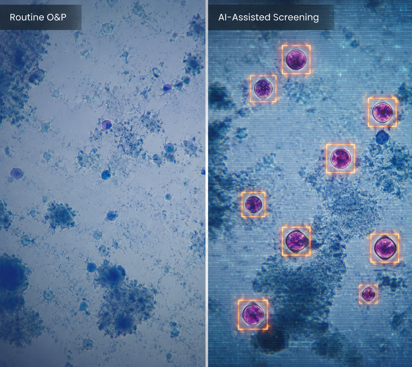
Where the routine O&P goes quiet
The miss is not mysterious to anyone who has had to make small organisms visible in stool. Routine ova-and-parasite examinations commonly use trichrome stain for permanent preparations. Cyclospora oocyst walls do not take up routine trichrome stain well, so the organism can remain faint or overlooked in a field crowded with stool debris. The CDC’s July 2026 health advisory states the operational consequence plainly: “routine ova and parasite examinations might not reliably detect the parasite,” and clinicians should specifically request Cyclospora testing.[2]
A single negative stool specimen also has limits because symptomatic patients may shed oocysts intermittently.[2] That matters at the callback stage. If the patient’s diarrhea keeps relapsing and the only test in the chart is a nonspecific O&P, the negative result has answered a narrower question than the clinician may think it has answered.
When Cyclospora is specifically requested, the laboratory can use methods designed for it. CDC identifies modified acid-fast staining as the best single method; modified safranin staining and ultraviolet fluorescence microscopy can also be used, and multiplex PCR panels that include Cyclospora are an alternative when available.[2] Those details are not academic. They determine whether a low-volume, intermittently shed organism is being actively sought or merely passing through a workflow built for a broader parasite screen.
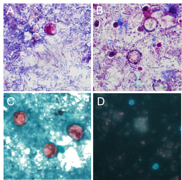
Why the 2026 outbreak exposes the diagnostic gap
The 2026 multistate outbreak turned this familiar weak point into a volume problem. As of mid-July 2026, CDC reported 1,645 laboratory-confirmed domestic cases, more than 5,100 probable cases, and 141 hospitalizations across 34 states in an outbreak linked to shredded iceberg lettuce served at Taco Bell.[3] By the comparable point in 2025, CDC surveillance had recorded 249 cases.[4]
Those numbers are not a reason to diagnose Cyclospora by news cycle. They are a reason to stop treating a routine O&P as if it closes the file. In a patient with compatible symptoms, exposure history, or residence in an affected area, the order has to tell the laboratory what is being considered.
| Clinical or laboratory issue | Why it matters for Cyclospora |
|---|---|
| Watery diarrhea that persists or relapses | The syndrome can continue for weeks to more than 1 month if untreated. |
| One negative stool specimen | Intermittent shedding means one specimen may not exclude infection. |
| Routine trichrome-based O&P | Cyclospora oocysts do not reliably take up routine trichrome stain. |
| Specific Cyclospora request | The lab can apply modified acid-fast stain, modified safranin, UV autofluorescence, or an appropriate PCR panel. |
What AI is doing in a real parasitology workflow
The useful AI story here is not a chatbot inferring an outbreak from vague gastrointestinal complaints. It is computer vision at the stool bench: digitized whole-slide images, a convolutional neural network that flags suspicious objects, and a trained technologist who decides what is real.
ARUP Laboratories began deploying AI for parasite screening in 2019, using a Techcyte-developed convolutional neural network to review whole-slide images and highlight suspicious organisms for technologist verification.[5] The system has been used across trichrome preparations since 2019, modified acid-fast preparations since 2021, and wet-mount preparations since 2025, with slide analysis taking approximately 2 minutes per slide.[5][6]
That placement in the workflow is the important part. AI does not turn a routine O&P into a definitive Cyclospora diagnosis. It makes candidate organisms harder to miss once the right preparation is in front of the system. In ARUP’s deployment, the algorithm flags; the technologist reviews; the laboratory reports.
During the 2026 outbreak surge, that distinction became practical rather than philosophical. ARUP reported an approximately 200% increase in testing volume and about 50 positive Cyclospora identifications per day from specimens submitted nationwide.[5] In that setting, the value of AI is not that it replaces parasitology expertise. It gives that expertise a narrower, highlighted search field when the number of slides rises.
ARUP has described validation studies showing significantly improved sensitivity compared with human-only analysis across trichrome, modified acid-fast, and wet-mount preparations, and a 2025 Journal of Clinical Microbiology publication validated AI-assisted wet-mount detection.[5][6] The publicly available summaries do not provide a full sensitivity-and-specificity table for every use case, so “improved sensitivity” should not be read as a portable performance number that applies to every laboratory, slide type, or specimen mix.
Screening is not confirmation
This is where language matters. AI-assisted microscopy is a screening aid inside a diagnostic process. It can reduce the chance that a suspicious 8-10 micrometer object is ignored on a busy day. It can help a reference laboratory maintain attention when testing volume climbs. It cannot supply the clinical suspicion that led to the right test order, and it cannot make a poorly matched protocol adequate after the fact.
The same caution applies to molecular testing. Multiplex PCR panels that include Cyclospora can be useful, but CDC notes that they are an alternative rather than a universally available default.[2] If a local panel does not include Cyclospora, or if the ordering pathway hides what organisms are actually covered, the clinician can still end up with a negative-looking result that does not answer the right question.
Treatment is also not the diagnostic center of gravity here, but it explains why delay matters. CDC identifies trimethoprim-sulfamethoxazole as treatment for cyclosporiasis.[1] A patient whose illness is allowed to cycle through “viral gastroenteritis,” “negative O&P,” and another week of symptoms has lost time because the diagnostic request was too general.
The produce-safety AI work is a different question
There is also AI work upstream of the clinic. University of Tennessee researchers, funded through the Center for Produce Safety, built a machine-learning system for high-throughput Cyclospora oocyst identification that reached 95% confidence in identifying oocysts, and the group is training the system to distinguish sporulated from non-sporulated oocysts.[7]
That is worth watching because sporulation status affects whether a positive environmental or produce sample suggests infectious risk. It is not the same evidence as a deployed clinical diagnostic workflow. A 95% confidence figure in a produce-safety project should not be borrowed to describe performance at a clinical stool bench.
What to order when Cyclospora fits
For the clinician, the practical move is simple and easy to miss: do not write only “O&P” when the syndrome or exposure history makes Cyclospora plausible. Specifically request Cyclospora testing. If using a molecular gastrointestinal panel, confirm that Cyclospora is included. If microscopy is used, understand whether the laboratory performs modified acid-fast staining, modified safranin staining, UV autofluorescence microscopy, or an AI-assisted workflow that still ends in human verification.
AI helps close the detection gap by making suspicious organisms harder to overlook, particularly when outbreak volume stresses the bench. It does not remove the need for a clinician to suspect Cyclospora, for the laboratory to use an appropriate method, or for a human reviewer to decide whether the highlighted object is truly the parasite.
References
- Clinical Overview of Cyclosporiasis, CDC.
- HAN Archive - 00531, CDC Health Alert Network, July 14, 2026.
- Investigation of Cyclosporiasis Outbreak: July 2026, CDC.
- Cyclosporiasis Surveillance, CDC.
- Cyclosporiasis Cases Surge; ARUP’s AI-Enhanced Testing Aids Diagnosis, ARUP Laboratories, July 13, 2026.
- AI Takes on Cyclospora Outbreak, BankInfoSecurity, July 17, 2026.
- AI used to speed up detection of Cyclospora in CPS project, Produce Processing.
Comments
Join the discussion with an anonymous comment.