
Search for ai in medical diagnosis examples and the field can look deceptively tidy: an algorithm reads an image, flags a disease, and helps a clinician decide faster. The deployed reality is less tidy and more useful. By Q1 2026, the FDA’s public list included more than 1,200 AI-enabled medical devices, but the category is heavily shaped by imaging; one 2025 analysis of 1,016 FDA authorizations through September 2024 found that 84.4% used imaging data, and radiology accounted for 76% of authorizations.[1][2]
That imbalance matters. It explains why many discussions of diagnostic AI sound like radiology discussions with other specialties attached later. A stroke triage algorithm that moves a CT angiogram into a stroke team’s line of sight is not the same clinical bet as an autonomous diabetic retinopathy device in primary care, an EHR sepsis model, a computational pathology reader, or an ECG-AI product. They may all sit under the phrase “AI diagnosis,” but they do not ask clinicians to trust the same thing.
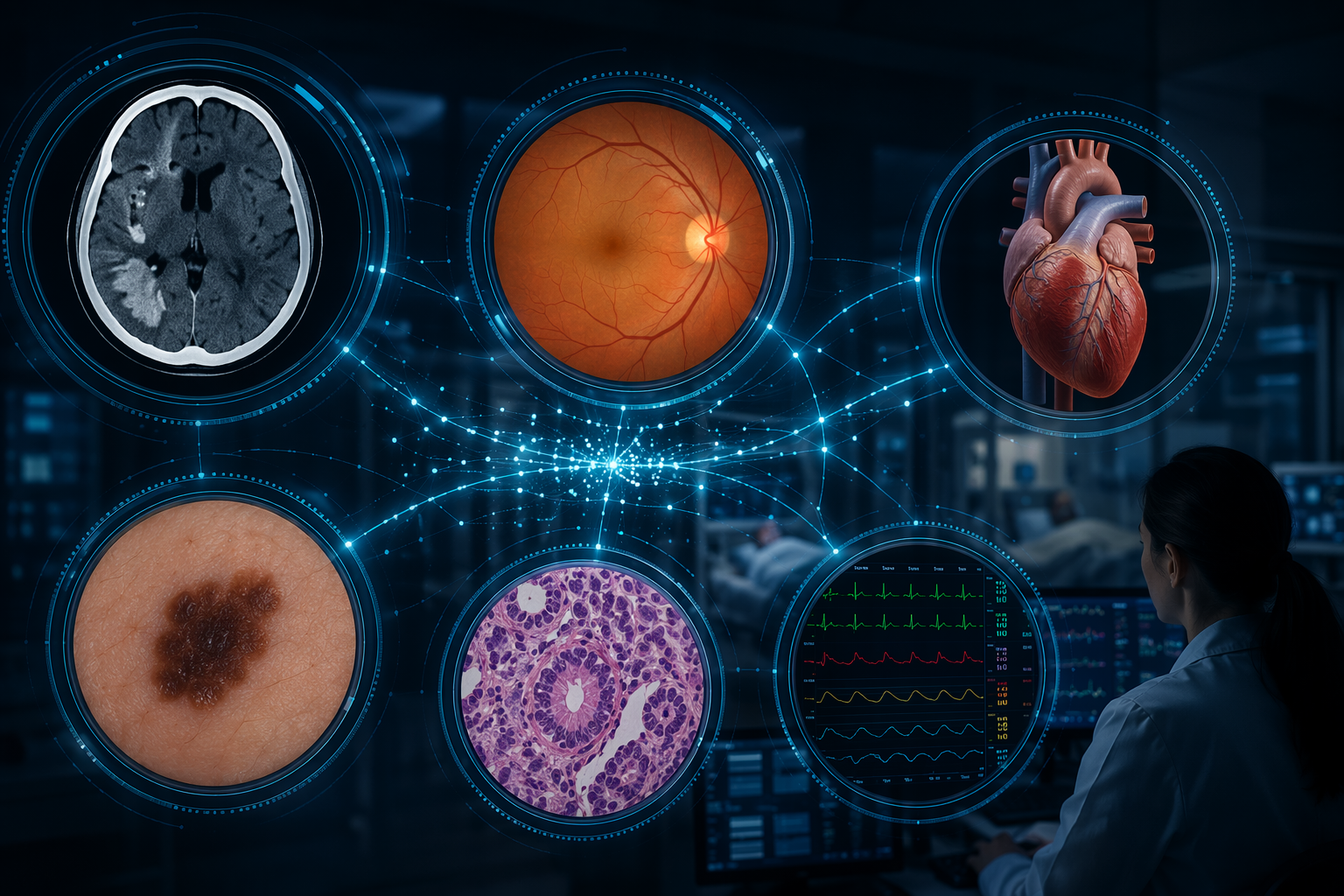
The examples below are limited to systems that are deployed or commercially used in clinical settings, have FDA authorization, FDA-listed regulatory status, or published clinical evidence in the cited sources, and have traceable documentation. That still leaves a mixed catalogue. Some products are workflow triage tools. Some are autonomous or near-autonomous diagnostic systems. Some are decision-support models whose value depends as much on alert routing and clinician response as on model discrimination.
A Scan-Level Catalogue Of 10 Deployed AI Diagnosis Systems
| System | Specialty | Clinical task | Regulatory pathway or status | Evidence tier | Deployment stage | Known limitation |
|---|---|---|---|---|---|---|
| Brainomix 360 | Stroke / radiology | Automated CT/MRI analysis and stroke team notification | FDA-authorized imaging AI device category | Qualitative workflow evidence plus regulatory documentation | Used in stroke networks | Acts before full clinician verification, so workflow design affects safety |
| Rapid LVO | Stroke / radiology | Large-vessel occlusion detection and alerting | FDA-authorized imaging AI device category | Workflow evidence and FDA device documentation | Used in acute stroke pathways | Triage benefit depends on who receives and acts on the alert |
| Viz LVO | Stroke / radiology | Suspected large-vessel occlusion detection and team activation | FDA-authorized imaging AI device category | Workflow evidence and FDA device documentation | Used in hospital stroke systems | False positives and false negatives have different operational costs |
| Epic Sepsis Model v2 | Sepsis / inpatient medicine | EHR-based prediction of sepsis risk from clinical data | Clinical decision-support AI deployed through EHR infrastructure | Multicenter prospective validation | Deployed in health-system settings including UPMC | Alert performance does not guarantee clinician action or outcome improvement |
| TREWS, Bayesian Health | Sepsis / inpatient medicine | Early warning from labs, vitals, and clinical documentation | Clinical decision-support AI deployed in hospitals | Published deployment and validation evidence | Deployed across Cleveland Clinic hospitals | Generalizability and response protocols remain central |
| IDx-DR / LumineticsCore | Ophthalmology / primary care | Autonomous detection of more-than-mild diabetic retinopathy | FDA De Novo | Pivotal trial and post-market evidence | Used at point of care without specialist interpretation | Autonomy is limited to the intended-use population and image-quality requirements |
| HeartFlow FFRct | Cardiology | Noninvasive fractional flow reserve analysis from coronary CT | FDA-cleared device documentation | Clinical studies plus regulatory documentation | Used after coronary CT angiography | It assesses lesion physiology from CT data, not every cause of chest pain |
| Anumana ECG-AI | Cardiology | ECG-based AI analysis for cardiac disease signals | FDA-cleared or FDA-authorized ECG-AI category | Regulatory documentation and clinical studies | Used in cardiac diagnostic workflows | ECG signal detection still requires clinical context and downstream confirmation |
| DermaSensor | Dermatology / primary care | Handheld AI spectroscopy assessment of skin lesions | FDA-authorized device documentation | Regulatory evidence and device studies | Used as a point-of-care lesion assessment aid | Evidence is narrower than a population-wide melanoma screening claim |
| Paige Prostate | Pathology | AI-assisted detection of prostate cancer on digitized pathology slides | FDA-authorized computational pathology device | Regulatory evidence and pathology studies | Used in digital pathology workflows | Performance depends on slide preparation, scanner workflow, and pathologist review |
This table is deliberately uneven. The systems are not ranked by excitement or model sophistication. They differ in what they touch: a queue, a specialist interpretation, a bedside alert, a primary-care screening visit, or a pathology review. The practical question is not whether the model is “AI,” but whether the clinical system around it makes the model’s output usable and safe.
Stroke Triage: AI As An Automated First Reader
Stroke triage is one of the clearest examples of diagnostic AI changing where attention goes. Brainomix 360, Rapid LVO, and Viz LVO are not replacing the stroke physician or neuroradiologist. Their more immediate role is to analyze CT or MRI data, identify suspected time-critical findings such as large-vessel occlusion, and distribute alerts to the stroke team quickly enough to change the order in which people look, call, and prepare.
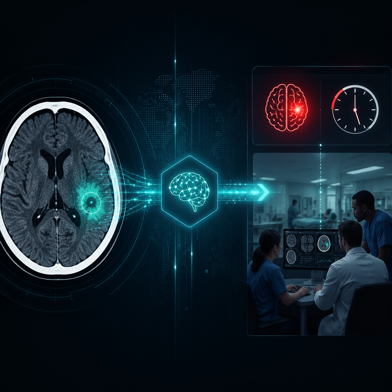
A five-year qualitative study of AI implementation at three UK stroke hubs described these systems as automated first readers embedded in stroke workflows, with AI results distributed to clinicians within minutes and sometimes triggering treatment pathways before full clinician verification.[3] That is exactly why the category is clinically interesting. The diagnostic act is not only image classification; it is the movement of a possible occlusion out of the ordinary reporting queue and into a team’s immediate field of action.
The same feature creates the hazard. A false-positive LVO alert can pull a stroke team into a pathway that consumes scarce attention. A false negative can leave a patient in the usual queue when minutes matter. A delayed alert, a phone that no one carries, or an unclear escalation protocol can erase the model’s theoretical advantage. Procurement slides often emphasize sensitivity, specificity, or time-to-notification; the clinician living with the system also needs to know who is notified, who verifies, how disagreement is handled, and whether the alert changes transfer or thrombectomy decisions.
Brainomix, RapidAI, and Viz.ai therefore belong in the same operational family but not in a single undifferentiated “AI detects stroke” bucket. Their value depends on a chain: scan acquisition, algorithm processing, image availability, alert delivery, clinician review, and treatment activation. Weakness anywhere in that chain becomes part of the diagnostic system, even if the model itself performs well in a validation dataset.
Sepsis Prediction: The Alert Is Only The Beginning
Sepsis AI feels different from stroke triage because there is no single image to point at and no clean moment when the model has “seen” the disease. Systems such as TREWS from Bayesian Health and the Epic Sepsis Model use EHR data streams: laboratory results, vital signs, structured clinical data, and, in some deployments, clinical notes. The aim is to identify patients at risk hours before conventional recognition, often in a noisy inpatient environment where nurses and physicians are already managing competing alerts.
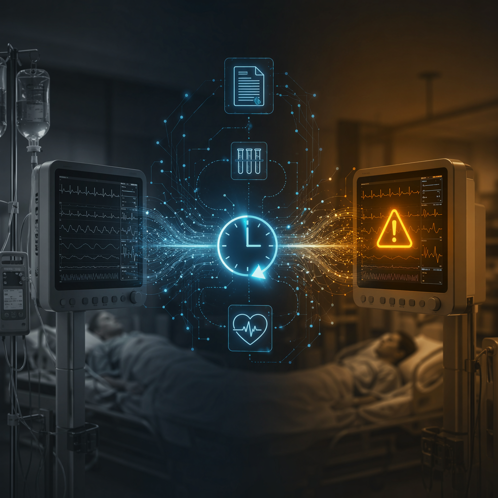
The strongest evidence anchor in this group is the 2026 multicenter prospective validation of Epic Sepsis Model v2, which evaluated 227,091 inpatient encounters.[4] That scale matters because sepsis prediction can look convincing in retrospective development work and then disappoint once alert volume, local practice patterns, missing data, and clinician behavior enter the system. A prospective validation does not settle every question, but it moves the conversation closer to the conditions under which the alert actually appears.
TREWS deserves attention for the same reason: it is not just a model in a paper, but an early-warning system deployed across Cleveland Clinic hospitals. Its clinical claim is not that it diagnoses sepsis the way a culture result identifies an organism. It surfaces risk earlier, so the important downstream questions are practical: does the alert reach someone with authority to act, does it trigger assessment rather than reflexive treatment, and does the institution monitor alert burden as carefully as model performance?
Sepsis is also where the phrase “AI diagnosis” can be most misleading. These systems are often prediction and early-warning tools rather than definitive diagnostic devices. A higher risk score can prompt review, lactate testing, cultures, antibiotics, fluids, or closer monitoring, depending on local protocols and patient context. The model does not bear the consequence of over-treatment, missed deterioration, antibiotic exposure, or alarm fatigue. The bedside team does.
Autonomous Diabetic Retinopathy Screening Is A Different Category
IDx-DR, now marketed as LumineticsCore by Digital Diagnostics, should not be casually grouped with triage tools. It was the first FDA-authorized autonomous AI diagnostic device, cleared through the De Novo pathway for detection of more-than-mild diabetic retinopathy in eligible adults with diabetes.[5] Its importance lies in the word autonomous: the device can produce a screening result in a primary-care setting without a specialist interpreting the retinal images at the point of care.
That changes the clinical setting. Instead of asking an ophthalmologist to look at another image faster, the system moves screening into a visit where the patient may already be seeing a primary-care clinician or diabetes care team. The intended benefit is not an abstract improvement in diagnostic intelligence; it is fewer missed opportunities for diabetic eye screening among patients who may not complete a separate ophthalmology referral.
The boundaries matter just as much. Autonomous does not mean universal. The device’s indication defines who can be screened, what image quality is acceptable, what disease threshold is being assessed, and when referral is required. For a deeper evidence discussion of the pivotal and post-market data, see the site’s focused review of IDx-DR and LumineticsCore clinical evidence.
Cardiology Systems: CT Physiology, ECG Signals, And Heart Sounds
HeartFlow FFRct is one of the more established cardiology examples because it operates at a specific decision point: after coronary CT angiography, it estimates fractional flow reserve noninvasively to help assess whether a coronary lesion is likely to be functionally significant. That is a narrower and more useful claim than “AI diagnoses heart disease.” It helps clinicians decide whether anatomy seen on CT is likely to correspond to flow-limiting disease, potentially reducing unnecessary invasive angiography in selected patients, based on the clinical studies and FDA documentation cited in the available materials.[1]
Anumana ECG-AI and Eko Health sit in a different part of cardiology. Anumana analyzes ECG data for cardiac disease signals; Eko Health analyzes heart sounds and ECG-related inputs in auscultation workflows. These systems are appealing because ECGs and heart sounds are already routine, cheap, and widely distributed. The diagnostic boundary is also obvious: a signal that suggests disease is not the same as a complete cardiac diagnosis. It needs confirmation, clinical context, and a workflow for what happens when the signal is unexpected.
Cardiology AI therefore spans at least two different procurement questions. HeartFlow is tied to a CT-based pathway after an anatomic test has already been ordered. ECG-AI and auscultation AI aim to extract more diagnostic signal from front-line tests. The first asks whether a noninvasive analysis can refine an invasive decision. The second asks whether a common signal can safely change who gets further evaluation.
Dermatology And Pathology: Similar Labels, Different Review Burdens
DermaSensor is a handheld device that uses spectroscopy with AI-based analysis to assess skin lesions. Its clinical attraction is the primary-care setting: a clinician facing an uncertain lesion can get an additional assessment before deciding whether to refer, biopsy, or monitor. The available regulatory evidence supports an authorized lesion-assessment aid, not a blanket claim that the device can replace dermatology expertise or population-level skin cancer screening.[1]
Paige Prostate is a different kind of diagnostic aid. It works inside computational pathology, helping identify prostate cancer on digitized slides. The FDA authorization of Paige Prostate is notable because pathology AI enters a high-liability interpretive workflow where the human pathologist remains central. The system can draw attention to suspicious regions, but the diagnostic report still depends on tissue quality, slide preparation, scanner performance, review behavior, and the pathologist’s judgment.[1]
Ibex Galen belongs near this discussion even though it is not one of the ten table entries above. Its breast, prostate, and gastric cancer detection products show how computational pathology is expanding beyond a single organ system in deployed settings. The important comparison is not whether one vendor’s interface looks more polished than another’s; it is whether the laboratory has validated the system with its scanners, staining variability, case mix, and review protocols. For more context on Paige’s position in this category, see the site’s Paige AI computational pathology profile.
What The Evidence Does Not Let You Assume
FDA authorization is a threshold, not a guarantee that a system will improve care in every hospital that buys it. The evidence base for clinical AI remains uneven. The Stanford-Harvard ARISE State of Clinical AI 2026 report, citing Bedi and colleagues, noted that nearly half of medical AI studies tested models on exam-style questions, only 5% used real patient data, and very few examined bias or fairness.[6] That critique is broader than the ten deployed systems discussed here, but it is a useful warning against treating the existence of many AI products as proof of mature clinical evidence.
The type of evidence also differs by system. A multicenter prospective validation of an EHR sepsis model answers different questions than a De Novo authorization for autonomous retinal screening, a qualitative study of stroke hub workflow, or FDA documentation for a point-of-care lesion device. A hospital choosing among these products should not ask for one generic “AI accuracy” number. It should ask what the device was authorized to do, where it was validated, what patient population was represented, what changed in the workflow, and what failure mode would matter most locally.
Automation bias is the quiet risk across categories. Studies summarized in the available materials found that clinicians sometimes followed incorrect AI recommendations even when errors were detectable.[6] The risk is not limited to inexperienced users or dramatic black-box systems. A plausible alert arriving at the right moment, in the right interface, with the right institutional blessing can become harder to question than a standalone model output.
That is why the most useful reading of these AI diagnosis examples is specific rather than celebratory. Brainomix 360, Rapid LVO, and Viz LVO are time-sensitive stroke triage tools. Epic Sepsis Model v2 and TREWS are inpatient early-warning systems whose performance depends on alert design and response. LumineticsCore is an autonomous retinal screening device within a defined intended use. HeartFlow FFRct, Anumana ECG-AI, Eko Health, DermaSensor, Paige Prostate, and related pathology systems each sit inside a particular diagnostic chain.
Taken together, they show that AI diagnosis is no longer speculative. They do not show that diagnostic AI has become a uniform clinical capability. Each system should be read as a regulated tool with a specific intended use, evidence base, deployment setting, and automation-bias risk.
References
- Artificial Intelligence-Enabled Medical Devices — U.S. Food and Drug Administration.
- How AI is used in FDA-authorized medical devices: a taxonomy across 1,016 authorizations — Nature npj Digital Medicine, 2025.
- D'Adderio & Bates study on AI in stroke workflow — Nature npj Digital Medicine, 2025.
- Wong et al. multicenter prospective validation of Epic Sepsis Model v2 — JAMA Network Open, 2026.
- FDA permits marketing of artificial intelligence-based device to detect certain diabetes-related eye problems — U.S. Food and Drug Administration, 2018.
- State of Clinical AI 2026 — Stanford-Harvard ARISE, 2026.
Comments
Join the discussion with an anonymous comment.