
By mid-July 2026, ARUP Laboratories was not dealing with a modest seasonal bump. Its Cyclospora testing volume had risen by 200%, and the laboratory reported about 50 positive cases per day while using an AI-enhanced microscopy platform to help screen slides in roughly two minutes each.[1] That is the practical setting in which AI-assisted Cyclospora testing becomes worth discussing: not as an abstract automation story, and not as patient-level treatment advice, but as a way to keep low-burden positives from disappearing inside a high-volume microscopy queue.
The outbreak pressure was real. CDC’s July 2026 health advisory reported 1,644 laboratory-confirmed cyclosporiasis cases, 94 hospitalizations, a median patient age of 44 years, and 56% female patients; the agency also identified iceberg lettuce as the suspected vehicle in the multistate outbreak.[2] Those numbers explain why reference laboratories were seeing more specimens. They do not, by themselves, explain why the detection problem is so easy to underestimate.
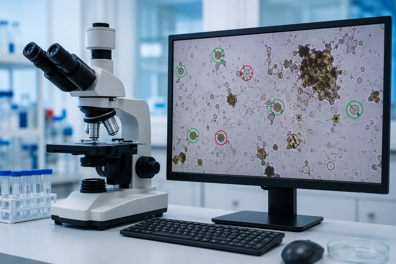
Cyclospora is a bad organism to look for when a bench is already saturated. CDC warned clinicians and laboratories that routine stool ova-and-parasite examinations may miss Cyclospora unless the laboratory is specifically asked to test for it.[2] ASM’s 2026 guidance likewise focuses on deliberate detection and reporting rather than assuming routine parasitology workflows will reliably surface cases.[3] The earlier diagnostic-gap problem is familiar: patients may have compatible illness, but the specimen, stain, organism burden, and test order do not line up cleanly. For a fuller discussion of those clinical gaps, see how Cyclospora symptoms from iceberg lettuce evade diagnosis.
The weak point is the visual search, not the technologist
A false negative in Cyclospora testing rarely announces itself as a dramatic laboratory failure. It looks more ordinary: a slide with too few organisms, a field full of distracting debris, an oocyst that does not take up stain cleanly, a technologist who has already reviewed hundreds of similar fields, and a patient whose result returns negative even though the clinical course keeps pulling them back into care.
ARUP’s own description of the problem is appropriately microscopic. Cyclospora oocysts may be shed intermittently and in low numbers, and routine trichrome staining is not reliable for this organism because the stain does not penetrate the oocyst wall well.[1] That combination matters more than raw specimen volume. A laboratory can add people, extend shifts, or batch work more aggressively, but none of those measures changes the fact that the positive object may be rare on the slide.
This is where fatigue becomes a diagnostic variable. The issue is not that trained microscopists lack skill. It is that the task asks human attention to remain uniformly sharp across repetitive, low-prevalence visual fields. During an outbreak, the workload increases at the same time that the consequence of a missed positive becomes more visible to clinicians, infection prevention teams, and public health surveillance.
What ARUP changed in the slide review
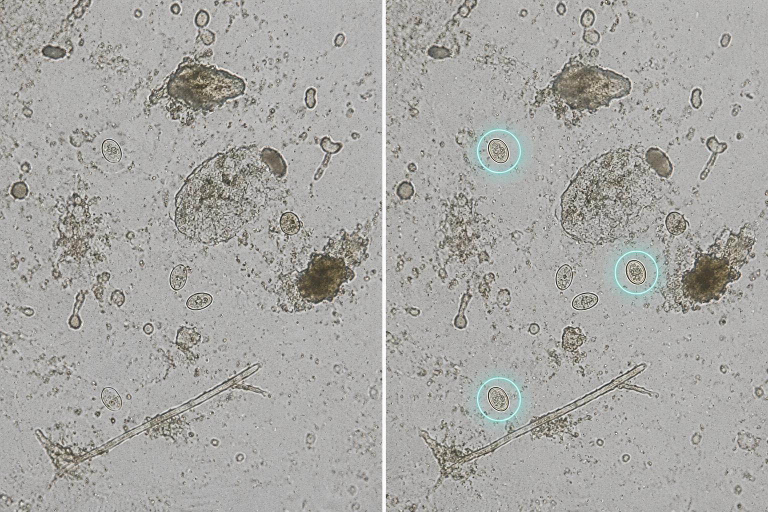
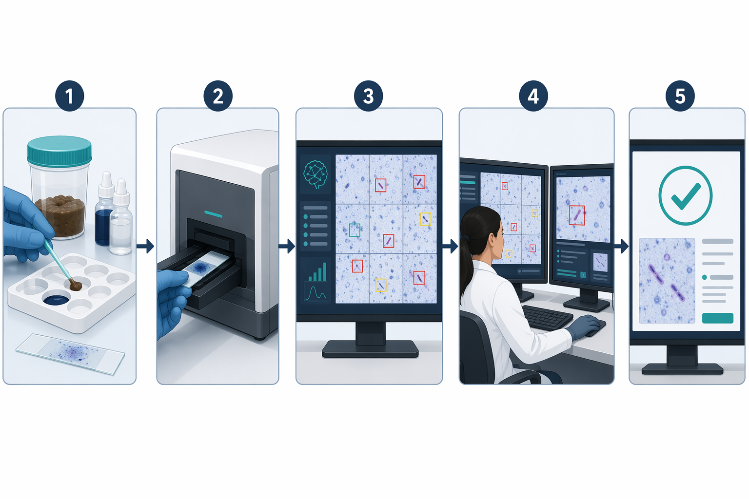
ARUP’s CNN-based workflow changes the first pass through the slide. Instead of asking a technologist to perform an unaided search from start to finish, the system scans the prepared slide, analyzes the digital image, and highlights suspicious objects for a human reviewer to confirm.[1] The decision still returns to the laboratory professional. The important shift is that the software absorbs the most vigilance-sensitive part of the work: finding candidate organisms in a field where the positive signal may be sparse.
| Workflow point | What changes with CNN-assisted screening |
|---|---|
| Specimen and staining | The laboratory still depends on an appropriate Cyclospora testing route rather than assuming routine stool testing is sufficient. |
| Slide imaging | The slide is digitized so the model can evaluate microscopy fields consistently at high volume. |
| CNN screening | The model flags objects with suspicious morphology instead of requiring the reviewer to find every candidate unaided. |
| Technologist review | A trained technologist confirms or rejects highlighted candidates within the laboratory workflow. |
| Resulting failure mode | The process moves from open-ended visual search toward assisted adjudication, while retaining human confirmation. |
The distinction is not cosmetic. A stand-alone diagnostic claim would require a different level of public evidence than ARUP’s currently available materials provide. What is documented is a deployed screening-support workflow: convolutional neural network analysis, object highlighting, and technologist confirmation, used at outbreak scale in a reference-lab setting.[1] That is still clinically meaningful. It gives the reviewer a narrower and more reproducible task.
ARUP’s timeline also makes the deployment more credible than a sudden outbreak-season technology announcement. The laboratory reported introducing AI for trichrome slide screening in 2019, expanding the platform to Cyclospora detection with modified acid-fast staining in 2021, adding wet mount screening in 2025, and then applying the system during the 2026 Cyclospora surge.[1] That sequence suggests an accumulation of laboratory process knowledge: staining, scanning, annotation, review, quality control, and user trust all had to mature before the outbreak tested the system.
Why Cyclospora is a useful stress test for laboratory AI
Some AI laboratory applications promise speed where speed is already the dominant operational metric. Cyclospora asks a harder question: can software reduce missed positives when the target is uncommon on the slide, inconsistently shed, and not reliably detected by routine methods? ARUP says its AI-assisted approach improves sensitivity compared with human-only analysis, but the public press release does not provide the exact sensitivity and specificity values needed to evaluate that claim independently.[1]
That missing detail matters. A 200% increase in testing volume and about 50 positives per day show operational load; they do not quantify diagnostic performance.[1] The CDC advisory’s case count and hospitalization count show outbreak scale; they do not prove that any one laboratory method is superior.[2] The credible argument for CNN screening rests on the match between the tool and the failure mode. CNNs are well suited to repeated image classification tasks, while technologists are essential for contextual review, stain-quality judgment, and final laboratory interpretation.
In practical terms, the model is not being asked to understand diarrhea, exposure history, treatment response, or outbreak epidemiology. It is being asked to help find objects that look suspicious enough to deserve expert attention. That narrower task is exactly why the workflow is plausible. It does not pretend that diagnosis begins and ends with an algorithm; it places the algorithm at the point where repetitive visual search is most vulnerable.
Diagnosis still controls treatment access
Treatment belongs in this discussion only because detection controls whether the patient gets managed for the right disease. The CDC advisory was directed at clinicians and public health partners during an ongoing outbreak, and it emphasized testing and reporting as part of case identification.[2] If routine stool testing misses Cyclospora, a patient may remain in the system with persistent symptoms while the laboratory record appears reassuring.
That is the clinical consequence of a microscopy miss. It is not merely a delayed result; it is a wrong negative that can redirect the next clinical decision. AI-assisted screening has value here because it may help preserve detection quality during precisely the period when volume makes unaided review more fragile. It should not be framed as treatment technology. It is diagnostic infrastructure that can make appropriate treatment and public health reporting more likely to begin from the correct result.
What makes the ARUP model hard to copy
The tempting conclusion is that other laboratories should simply adopt AI microscopy for Cyclospora. The more careful conclusion is that ARUP’s model depends on conditions that many laboratories may not have in place. A reference laboratory with substantial parasitology volume can generate and review enough material to support model development, validation, monitoring, and workflow refinement. A smaller hospital laboratory may see too few positives to build the same local evidence base.
- Validated staining and slide-preparation workflows, because the model can only evaluate the image quality it receives.
- Annotated training and validation material that represents the organism, stain variation, debris, artifacts, and low-density positives expected in real specimens.
- A quality-control process that tracks model behavior over time instead of assuming performance remains stable after deployment.
- Technologist confirmation, with the system treated as a screening aid unless regulatory status and validation evidence support a broader claim.
- Enough test volume to justify the scanner, software, training, review process, and oversight burden.
The public record still leaves several adoption questions unresolved. ARUP’s announcement refers to validation work and improved sensitivity, but it does not publish the exact performance metrics in the announcement itself.[1] It also does not provide enough detail on the number of annotated images used, how broadly the training set captured staining variation, how poor-quality slides are handled, or whether the platform’s regulatory status is best understood as a screening aid rather than a stand-alone diagnostic device.
Those are not objections to AI screening. They are the questions that determine whether a successful reference-lab deployment can travel. A laboratory director does not need a promise that the algorithm is intelligent; they need to know what specimens it was trained on, which stain preparations it accepts, how discordant cases are reviewed, what happens when slide quality falls below threshold, and who signs out the result.
A better failure mode during an outbreak
ARUP’s 2026 experience is persuasive because the AI is embedded inside a laboratory process that already understands microscopy. The system did not remove staining decisions, specimen limitations, technologist judgment, or quality control. It changed the weakest part of the outbreak workflow: the expectation that a human reviewer can maintain perfect visual search performance while volume rises and organism burden remains low.
That is enough to make CNN-powered screening a serious model for Cyclospora detection at scale. It is not enough to make it automatically transferable to every laboratory. The strongest version of the approach remains tied to adequate volume, disciplined slide preparation, validated image analysis, transparent performance metrics, and human confirmation. Until the exact validation data, regulatory pathway, training-set requirements, and generalizability limits are clearer in the public record, ARUP’s deployment should be read as a strong reference-lab example rather than a plug-and-play answer for smaller testing sites.
References
- Cyclosporiasis Cases Surge; ARUP’s AI-Enhanced Testing Aids, ARUP Laboratories, July 13, 2026, https://www.aruplab.com/news/07-13-2026/cyclosporiasis-cases-surge-arups-ai-enhanced-testing-aids
- HAN Archive - 00531, Centers for Disease Control and Prevention, July 2026, https://www.cdc.gov/han/php/notices/han00531.html
- Cyclospora Detection and Reporting from Clinical Specimens, American Society for Microbiology, July 2026, https://asm.org/guideline/cyclospora-detection-and-reporting-from-clinical-s
Comments
Join the discussion with an anonymous comment.