
The most important fact about Google AI chips in medical applications is also the easiest one to blur: Google’s TPUs have helped produce several landmark medical AI results, but as of mid-2026 there is no FDA-cleared device filing identified in the available materials that explicitly names TPU or Edge TPU as the compute platform. That does not make the chips irrelevant. It means the evidence has to be read in layers: peer-reviewed clinical task performance, official regulatory records, then engineering or cloud infrastructure claims.
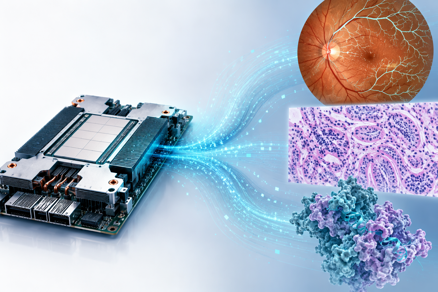
A TPU, or Tensor Processing Unit, is Google’s custom accelerator for machine learning workloads. In medicine, two quite different families matter. Cloud TPUs sit in datacenters and make it practical to train or run large vision, protein, and imaging models at scale. Edge TPU, sold through the Coral ecosystem, is a low-power inference chip intended for local, quantized models. The first category is mostly visible in research and cloud pipelines; the second is the one people imagine in bedside, microscope, clinic, or point-of-care devices. Those are not interchangeable claims.
Where the Clinical Evidence Is Strongest
The strongest bridge between TPUs and recognizable medical work is medical imaging: diabetic retinopathy screening, lymph node metastasis detection in breast cancer pathology, and lung cancer prediction from CT. These are not abstract benchmark tasks. They are the kinds of problems that put clinicians in front of images, triage queues, false negatives, and workflow bottlenecks.
The diabetic retinopathy work remains the cleanest entry point. Google describes a 2016 JAMA study in which a deep learning system was evaluated on more than 10,000 retinal fundus images and performed at a level comparable with board-certified ophthalmologists for detecting referable diabetic retinopathy.[1] The clinical importance is plain: screening programs need scalable image review, and ophthalmology capacity is not evenly distributed. The TPU question, however, is more delicate. Google’s broader imaging materials connect this line of work to Google-scale AI infrastructure, but the clinical result should still be read as evidence for the model’s diagnostic performance in that study setting, not as evidence that a TPU-based retinal screening device had been cleared or deployed.
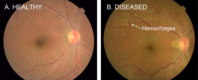
That distinction matters because a retinal screening algorithm can be scientifically impressive before it is operationally proven in a specific health system. A paper can show performance against expert grading; a hospital still has to know who acquires the image, what happens when image quality is poor, where the result appears in the record, which clinician is responsible for follow-up, and what device or cloud architecture is covered by regulatory and procurement documents.
The breast pathology work is technically beautiful for a different reason. Google’s LYmph Node Assistant, or LYNA, was built to detect metastatic breast cancer in lymph node slides. In the 2018 Archives of Pathology & Laboratory Medicine study described by Google Research, LYNA achieved 99% slide-level accuracy for detecting metastatic cancer. In a proof-of-concept workflow involving 6 board-certified pathologists, use of the system reduced review time for micrometastases from about 2 minutes to about 1 minute.[2]
Those are the kinds of numbers that should make a pathology department pay attention. The result combines a diagnostic endpoint with a workflow endpoint, and the workflow endpoint is not cosmetic: time spent on small foci of disease is exactly where fatigue and opportunity cost accumulate. But the caveat belongs in the same breath. The pathologist component was a proof-of-concept involving 6 clinicians, not a broad live deployment across routine pathology operations. It supports the idea that AI assistance could reduce review burden under study conditions; it does not establish that an Edge TPU microscope or any named TPU-powered clinical device became the standard implementation.
The lung cancer CT study extends the pattern to volumetric imaging. Google Research describes a Nature Medicine 2019 system for lung cancer prediction using 3D volumetric modeling, with an AUC of 94.4% on a 6,716-case validation set.[3] Here the TPU relevance is easy to understand architecturally: 3D CT modeling is computationally heavier than many 2D image classification tasks, and large accelerators make model development more practical. Still, the clinical paper’s performance result and the engineering infrastructure story are not the same evidentiary object.
| Clinical task | What the cited evidence supports | What it does not establish |
|---|---|---|
| Diabetic retinopathy screening | Deep learning performance comparable with board-certified ophthalmologists on more than 10,000 retinal fundus images | An FDA-cleared device filing that explicitly names TPU as the compute platform |
| Breast cancer lymph node metastasis detection | 99% slide-level accuracy and reduced review time in a 6-pathologist proof-of-concept workflow | Routine deployment across live pathology departments or dedicated Edge TPU clinical validation |
| Lung cancer CT prediction | AUC 94.4% on a 6,716-case validation set using 3D volumetric modeling | A procurement-ready TPU-based lung cancer product with explicit regulatory attribution |
This is where infrastructure becomes both important and easy to over-credit. Without large-scale compute, some of these experiments would have been slower, narrower, or less practical. Yet clinicians do not deploy accelerators; they deploy workflows, devices, software updates, support contracts, audit trails, and escalation paths. The chip can be essential to producing the model and still absent from the evidence needed to purchase or clear the clinical product.
AlphaFold Belongs in the Biomedical Column, Not the Bedside Device Column
AlphaFold is the case where TPU-enabled scale has most clearly changed biomedical research. Google reports that AlphaFold has predicted more than 200 million protein structures and has been cited in more than 20,000 publications; the model family was trained on TPU v3 and v4 pods.[4] That is not a marginal infrastructure footnote. Protein structure prediction at that scale changes what researchers can look up, screen, compare, and hypothesize before an experiment begins.
It would still be a mistake to describe AlphaFold as a direct clinical diagnostic deployment. Its medical value is upstream: drug discovery, molecular biology, target understanding, structural hypotheses, and research prioritization. A hospital does not diagnose pneumonia with AlphaFold. A laboratory or pharmaceutical team may use protein structure information to shape experiments that eventually influence therapeutic development.
That upstream position does not make the work less consequential. It simply places the evidence in the right lane. AlphaFold’s scale and scientific uptake are real; its relationship to patient care is mediated through research pipelines, not through a cleared bedside or imaging device.
Drug Discovery Claims Need a Shorter Leash
The Bayer-Google Cloud collaboration is a useful example of how quickly a plausible compute story can outrun available outcomes. Bayer announced in January 2023 that it would work with Google Cloud on high-performance computing for drug discovery, including density functional theory for protein-ligand modeling.[5] The direction is credible: quantum chemistry and molecular modeling are compute-intensive, and cloud TPU resources are naturally attractive for this class of work.
But an announced collaboration is not the same as a peer-reviewed therapeutic result. In the materials available for this article, no published peer-reviewed outcomes from that Bayer-Google Cloud TPU work were identified by mid-2026. The safest conclusion is narrow: the collaboration shows industry interest in using Google Cloud’s high-performance compute for drug discovery workloads, not demonstrated clinical effectiveness or drug development success attributable to TPUs.
Edge TPU Is Promising for Point-of-Care Medicine, but the Validation Gap Is Larger
Edge TPU is attractive for reasons that clinical teams can understand without memorizing chip roadmaps. A small local accelerator could run inference close to the scanner, microscope, camera, or clinic workstation. That may reduce latency, limit data movement, and preserve some functionality where network connectivity is unreliable. Coral ecosystem descriptions cite 4 TOPS at 2W, with a systolic-array matrix multiplication unit designed for quantized inference.[6]
Those specifications are relevant to medical device design, especially for low-power point-of-care settings. They are not clinical validation. The available materials do not identify dedicated peer-reviewed clinical studies showing that an Edge TPU-powered medical device improves diagnosis, triage, outcomes, or workflow in a validated care setting.
The Augmented Reality Microscope line of work is the most concrete reason not to dismiss the edge idea. Google’s publication describes an augmented microscope with real-time artificial intelligence integration for cancer diagnosis, and the project has been associated with on-device inference lineage and a 2023 Department of Defense collaboration.[7] The clinical image is compelling: a pathologist looks through a microscope and sees AI-generated cancer heatmaps in the field of view rather than waiting for a separate workstation or cloud round trip.
Still, the practical question is not whether the concept is elegant. It is whether a specific device, running a specific model on a specific compute platform, has been clinically validated and cleared for the intended use. For Edge TPU medical applications, the evidence base is thinner than the enthusiasm around local inference would suggest.
Cloud Platforms Can Support Medical AI Without Proving Clinical Adoption
Google Cloud’s medical imaging ecosystem also deserves careful placement. The Google Cloud Medical Imaging Suite, launched in October 2022, has been described as offering DICOM-compliant storage, annotation support, MONAI pipelines, and access to accelerator-backed cloud infrastructure.[8] That kind of platform can matter a great deal to imaging scientists and hospital AI teams trying to manage data, training, annotation, and deployment pipelines.
But platform availability is not the same as clinical use at scale. A cloud suite can lower the friction of building and testing medical imaging models; it does not by itself show that a TPU-backed model is safe, effective, reimbursed, maintained, integrated into clinical worklists, or accepted by the clinicians who must act on its output.
How to Read a TPU Medical Claim
For a hospital AI lead or procurement team, the useful question is not “Were TPUs involved?” The useful question is what the TPU involvement permits you to believe.
- If TPUs were used to train a model, the claim may support scalability of model development, not necessarily clinical deployment.
- If a peer-reviewed clinical paper reports diagnostic performance, the result applies to the study design, dataset, readers, and endpoint described.
- If a company blog identifies TPU infrastructure, treat it as useful engineering context, not a substitute for clinical validation.
- If a regulatory filing does not name the compute platform, do not infer TPU-based clearance from brand association alone.
- If an edge device is proposed for point-of-care use, ask for validation of the full device-model-workflow combination, not just chip specifications.
This evidence hierarchy is not hostile to Google’s hardware. Quite the opposite: it keeps the hardware contribution visible without letting it absorb credit that belongs to a clinical study, a regulatory submission, or a deployed workflow. TPUs have clearly mattered in producing important medical AI and biomedical research results. The remaining gap is translation: explicit regulatory attribution, real-world workflow evidence, and dedicated validation for Edge TPU devices have not caught up with the scale of the research achievements.
References
- Imaging and diagnostics, Google Health, https://health.google/imaging-and-diagnostics/
- Applying deep learning to metastatic breast cancer detection, Google Research Blog, https://research.google/blog/applying-deep-learning-to-metastatic-breast-cancer-detection/
- Healthcare, Google Research, https://research.google.com/teams/brain/healthcare/
- AI models advancing health research, Google Health, https://health.google/ai-models/
- Bayer to accelerate drug discovery with Google Cloud’s high-performance compute power, Bayer, January 2023, https://bayer.com/media/en-us/bayer-to-accelerate-drug-discovery-with-google-clouds-high-performance-compute-power/
- Google Coral for Edge AI: The Complete Guide, viso.ai, https://viso.ai/edge-ai/google-coral/
- An Augmented Reality Microscope with Real-time Artificial Intelligence Integration for Cancer Diagnosis, Google Research, https://research.google/pubs/an-augmented-reality-microscope-with-real-time-artificial-intelligence-integration-for-cancer-diagnosis/
- MONAI Drives Medical AI on Google Cloud with Medical Imaging Suite, NVIDIA Developer, October 2022, https://developer.nvidia.com/blog/monai-drives-medical-ai-on-google-cloud-with-medical-imaging-suite/
Comments
Join the discussion with an anonymous comment.