
AI pathology foundation model applications now cover more than slide classification. The clinically relevant map has at least five branches: cancer diagnosis and subtyping, biomarker prediction, prognosis and survival prediction, rare cancer retrieval, and automated report generation. The boundary is just as important as the breadth: through April 2026, only 7 whole-slide imaging AI algorithms had FDA authorization, and none were pathology foundation models cleared as clinical products.[1]
For readers who need the basic distinction between a foundation model and a narrow pathology classifier, the short version is available in this foundation models in healthcare glossary. The clinical question here is narrower: which uses have evidence that a pathology department, tumor board, or health system evaluator can actually inspect?
| Application category | Leading cited models or benchmarks | Strongest reported signal | Evidence maturity | Main caveat |
|---|---|---|---|---|
| Cancer diagnosis and subtyping | TITAN; CONCH; Virchow2; PathBench models | TITAN outperformed PRISM, GigaPath, and CHIEF across 14 morphological tasks and reported average gains of +8.4% on multiclass and +6.7% on binary subtyping over the next best model.[2] | Stronger research evidence; clearer task framing; multiple-model comparison available | Relative gains can obscure modest absolute performance and organ-level variation. |
| Biomarker prediction | TITAN; Virchow2; independent 2026 benchmark | The 2026 independent benchmark found Virchow2 leading in biomarker prediction across a 31-task evaluation of 19 foundation models.[3] | Stronger research evidence; especially useful as a hypothesis-generating or triage layer | Prediction is not the same as a clinically acceptable substitute for validated molecular or IHC testing. |
| Prognosis and survival prediction | TITAN; CONCH; PathBench models | TITAN included 6 survival tasks, and the 2026 benchmark found CONCH leading in prognosis.[2][3] | Promising but less mature | Survival endpoints are harder to validate because treatment, stage, follow-up, and institution-level care patterns influence outcomes. |
| Rare cancer retrieval and case-based reasoning | TITAN; zero-shot retrieval benchmark | TITAN reported +14.8% Accuracy@K for rare cancer retrieval and +30.8% on an external Japanese cohort over GigaPath, across 186 cancer types without fine-tuning.[2] | Clinically intuitive research use; attractive for slide search and comparison | Independent zero-shot retrieval results show wide organ-level variability, with top-5 macro-F1 around 40-42% overall, kidney at 68%, and lung at 21%.[4] |
| Automated report generation | Foundation-model and generative AI research direction | Pathology foundation models have been explored across broad clinical pathology tasks, including IHC scoring and transplant-related assessment.[5] | Early research direction | Report text creates a higher-risk failure mode because fluent language can conceal unsupported inferences. |
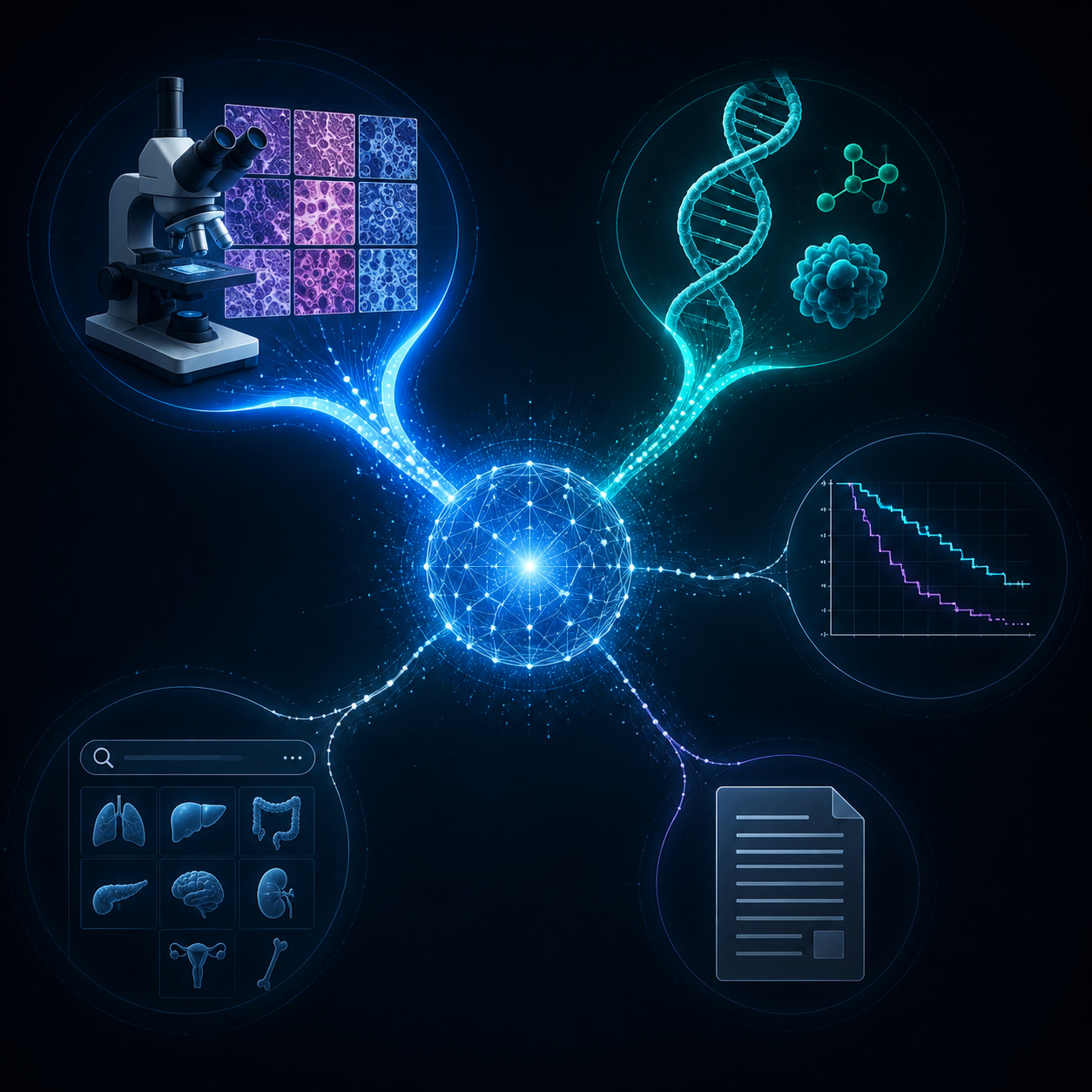
The evidence is broad, but not evenly clinical
TITAN is the obvious place to start because it shows why the field has momentum. In Nature Medicine, the model was evaluated across 14 morphological, 39 molecular, and 6 survival tasks, and it outperformed PRISM, GigaPath, and CHIEF across those task groups.[2] That is an unusually wide application map for pathology AI, and it points toward a practical idea: one slide-level representation could become a shared feature layer for classification, retrieval, molecular prediction, and survival modeling.
The independent 2026 Nature Biomedical Engineering benchmark is the necessary counterweight. It evaluated 19 foundation models across 31 tasks, using 6,818 patients and 9,528 slides, and found CONCH and Virchow2 tied as the top-performing models overall.[3] That result is helpful because it is not a developer-reported leaderboard for one model. It also makes the field look more sober: the top models reached only about 0.71 mean AUROC across the 31 tasks, which is not a clinical deployment number by itself.[3]
This distinction matters in a tumor board environment. A model that ranks first across many tasks may still be unsuitable for a specific organ, biomarker, or institution. A pathologist does not sign out an average AUROC. An oncologist does not order treatment from a relative gain. The useful question is which tasks have a clean enough target, enough external testing, and enough error visibility to be evaluated as near-clinical tools.
Cancer diagnosis and subtyping have the clearest evaluation shape
Diagnosis and subtyping are the strongest application category because the target is relatively familiar to pathology validation. A model predicts a class from a slide or region, and the result can be compared with a reference diagnosis. The evaluation still has traps, but the basic frame is recognizable: cancer type, subtype, grade-like morphology, or another histologic category.
TITAN’s subtyping results are therefore more clinically legible than some of the broader foundation-model claims. The paper reported average improvements of +8.4% on multiclass subtyping and +6.7% on binary subtyping over the next best model, while comparing against PRISM, GigaPath, and CHIEF.[2] Those are relative gains, not a license to deploy, but they are attached to tasks that pathology departments already know how to audit.
The 2026 independent benchmark gives the category a second, more comparative anchor. Across 31 tasks, CONCH led in morphology and prognosis, while Virchow2 led in biomarker prediction.[3] That split is useful because it argues against treating “best foundation model” as a universal label. A model can be stronger for morphology and weaker for biomarker inference, or vice versa, depending on architecture, training data, and downstream task design.
PathBench, an arXiv preprint, expands the comparison further: 19 pathology foundation models were evaluated on 15,888 whole-slide images from 8,549 patients across 10 hospitals, covering more than 64 diagnosis and prognosis tasks; Virchow2 and H-Optimus-1 were reported as the most effective overall.[6] Because it is a preprint, it should not be weighted the same way as peer-reviewed external validation, but it supports the same practical point: subtyping and diagnostic classification are where multi-model benchmarking is becoming dense enough to be useful.
The remaining problem is not whether these models can classify slides in research settings. They can. The problem is whether performance survives the exact case mix, scanner mix, staining practice, tissue preparation, and diagnostic boundary of the institution that would use them. For that question, an organ-level table usually matters more than the headline model name.
Biomarker prediction is compelling, but it must not be confused with testing
Biomarker prediction from H&E slides is one of the most attractive foundation-model applications because it promises to extract molecularly relevant signals from routine pathology material. In the best version of the workflow, the model does not replace a validated assay. It helps decide which cases deserve reflex testing, flags discordant cases for review, or supports research into morphology-genotype associations.
TITAN’s molecular task coverage is broad: 39 molecular tasks were included in the Nature Medicine evaluation.[2] The independent 2026 benchmark also separated biomarker performance from morphology and prognosis, and found Virchow2 leading in biomarker prediction.[3] That separation is exactly what clinical readers need. A biomarker model should be judged against biomarker tasks, not allowed to borrow credibility from strong cancer-subtyping results.
The strongest near-term role is probably enrichment rather than substitution. If a model helps a team identify cases more likely to be positive for a molecular feature, the consequence is additional review or testing. If it is used as a replacement for IHC, FISH, PCR, sequencing, or another validated method, the consequence is a direct change in diagnostic workup. Those are different clinical risks, and they require different evidence.
Mass General Brigham’s 2024 report illustrates how broad pathology foundation-model work is becoming outside oncology-only classification. The group described foundation models applied to more than 30 clinical tasks, including organ transplant rejection assessment, cardiac and renal allograft evaluation, and IHC scoring.[5] That breadth is encouraging, but IHC scoring and molecular inference still need task-specific validation. A single slide encoder does not make every downstream readout equally trustworthy.
Prognosis and survival prediction are clinically attractive and harder to validate
Survival prediction has obvious appeal. If a slide contains morphology associated with recurrence, progression, or mortality, a model could add information beyond a conventional diagnosis. TITAN included 6 survival tasks, and the independent benchmark found CONCH leading in prognosis.[2][3]
The validation burden is heavier than for subtyping. Survival reflects stage, treatment, comorbidities, access to care, follow-up length, and institution-specific practice patterns. A model may detect tumor biology, but it may also learn correlates of care delivery or cohort composition. That does not make prognosis modeling useless; it makes endpoint design and external validation central.
For clinical use, the question is not simply whether the model separates Kaplan-Meier curves in a study cohort. The question is whether the prediction changes a decision already made by a clinician, whether it adds information beyond established clinicopathologic variables, and whether its calibration holds outside the development environment. Current evidence supports research use and comparative benchmarking more strongly than direct prognostic deployment.
Rare cancer retrieval is the most intuitive application—and one of the easiest to overstate
Rare cancer retrieval has a different kind of appeal. A pathologist facing an unusual case does not necessarily need an autonomous diagnosis from a model. They may need a ranked set of similar slides, diagnoses, and annotations that help them decide what to consider next. That is a natural fit for a foundation model used as a slide-search layer.
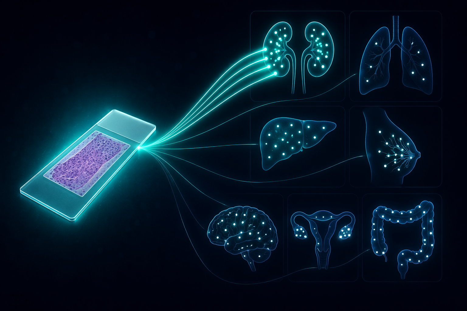
TITAN’s rare cancer retrieval result is one of its most clinically vivid findings. The model enabled off-the-shelf slide search across 186 cancer types without fine-tuning, and reported +14.8% Accuracy@K over GigaPath; on an external Japanese cohort, the reported gain was +30.8%.[2] That is the kind of use case that can be imagined in a real consult workflow: not “the model signs out the case,” but “the model retrieves candidate comparators for expert review.”
The independent zero-shot retrieval literature puts boundaries around that excitement. In Scientific Reports, an evaluation of 11,444 whole-slide images across 23 organs and 117 cancer subtypes found that leading foundation models achieved about 40-42% macro-averaged F1 for top-5 retrieval.[4] Organ-level variation was wide: kidney reached 68%, while lung was 21%.[4]
That spread is not a footnote. It is the difference between a retrieval system that may be helpful for one organ family and misleadingly noisy for another. In a clinical setting, every retrieved “similar case” creates work for someone: a pathologist must inspect it, decide whether the resemblance is meaningful, and prevent a visually plausible neighbor from becoming diagnostic anchoring.
Automated report generation remains the least ready clinical category
Report generation sits at the edge of the current evidence map. It is easy to see why it attracts attention: pathology reports are structured, repetitive in places, and clinically consequential. But generating diagnostic language is not the same as classifying a slide patch or retrieving a neighbor case.
The risk is not only that a generated report might be wrong. It might be wrong fluently. A generative system can produce text that sounds complete while omitting uncertainty, overstating a model output, or blending a true slide observation with an unsupported inference. That failure mode is harder to catch than a numeric biomarker probability sitting in a review queue.
For now, automated report generation is best treated as a research and documentation-support direction, not a mature clinical application of pathology foundation models. Drafting, summarization, and structured data extraction may prove useful, but they need separate evaluation from slide diagnosis. A model that retrieves similar rare tumors has not thereby earned permission to write a final surgical pathology report.
Why benchmark success has not become Tuesday-morning deployment
The regulatory boundary is straightforward. As of April 2026, FDA-authorized whole-slide imaging AI tools remained few, and the 7 authorized algorithms identified in the Innolitics review were not foundation models.[1] They were classified as in vitro diagnostics under 21 CFR Part 864 rather than as software as a medical device.[1] For more context on that pathway, see this overview of whole-slide imaging AI in clinical practice.
The evidence boundary is more subtle. Peer-reviewed developer studies such as TITAN are valuable, but independent external benchmarks carry a different kind of weight. Preprints such as PathBench can be useful for scanning the field, but their findings should remain provisional until peer review and replication. A health system evaluating a foundation model should separate those evidence levels before asking about integration, cost, or workflow.
- Use-case fit: subtyping, biomarker enrichment, retrieval, prognosis, and report drafting create different risks.
- External validation: performance should be shown outside the development institution and not only on pooled averages.
- Organ-level reporting: kidney-like retrieval performance and lung-like retrieval performance should not be averaged into a vague generalizability claim.
- Site-bias testing: evaluators should look for medical-center signatures, scanner effects, and staining patterns that could masquerade as diagnostic signal.
- Patch-scale limits: models built around small image patches may learn texture statistics that do not always align with diagnostically meaningful architecture.
- Regulatory status: research performance, even in a strong benchmark, is not FDA authorization.
This is where the current application map lands. Pathology foundation models already support meaningful research across cancer subtyping, biomarker prediction, prognosis, rare cancer retrieval, and early report-generation experiments. The strongest clinical evidence is in diagnosis/subtyping and biomarker prediction because those tasks have clearer labels, richer benchmarks, and more direct comparison across models. Survival modeling and zero-shot retrieval are promising but more variable. Automated report generation remains the most cautious category.
The useful distinction is not promise versus failure. It is research capability versus validated clinical deployment. On that distinction, pathology foundation models are no longer speculative as a research layer, but they are not yet cleared, general-purpose clinical instruments.
References
- AI/ML in Digital Pathology and the Software as an IVD Paradigm Snapshot, Innolitics, 2026.
- TITAN, Nature Medicine, 2025.
- Independent benchmark of pathology foundation models, Nature Biomedical Engineering, 2026.
- Zero-shot retrieval, Scientific Reports, 2025.
- Mass General Brigham researchers develop AI foundation models to advance pathology, Mass General Brigham, 2024.
- PathBench, arXiv, 2025.
Comments
Join the discussion with an anonymous comment.