
Before semi-automated offside technology could argue about soccer, it had to argue with bodies. At MIT Sports Lab, the early validation work found skeletal tracking data that was visibly impossible: skeletons flying above the ground, skeletons buried underground, limbs stretching from 30 cm to several meters. Those were not edge-case philosophical failures. They were bad inputs, and they were corrected before algorithm training began.[1]
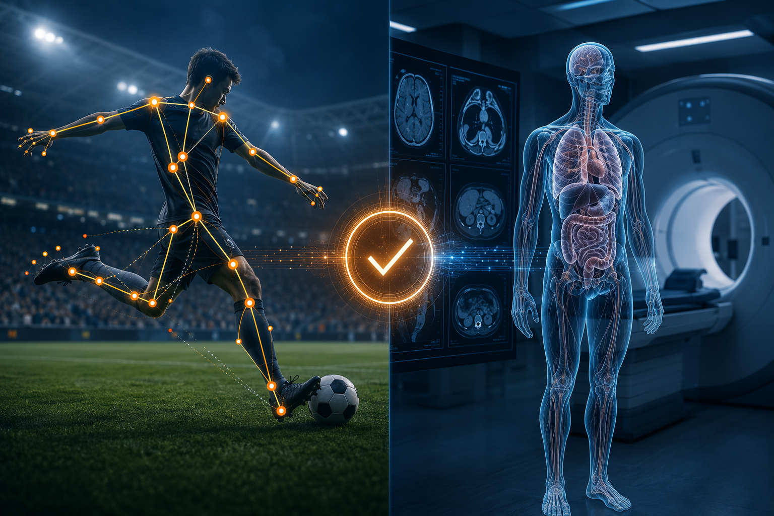
That is the useful entry point for clinicians looking at AI in semi-automated offside technology for soccer. The lesson is not that a stadium system and a radiology model solve the same problem. They do not. The useful lesson is that FIFA’s SAOT pathway treated computer vision validation as a sequence of separate obligations: prove that the incoming spatial data make physical sense, prove performance against an independent ground truth, then prove the system can survive live use before full authorization.
Medical imaging AI often gets discussed from the opposite direction. A paper reports a strong retrospective benchmark. A vendor demo shows clean overlays. A dashboard reports sensitivity, specificity, or AUC. Those numbers can matter, but they do not answer the first operational question: whether the system is receiving coherent data under the conditions in which it will actually be used.
The first validation layer was not accuracy
The MIT Sports Lab layer is easy to underestimate because it sounds less glamorous than an accuracy claim. It was a data integrity layer. The question was not yet whether SAOT could make the right offside recommendation. The question was whether the player-tracking representation could be trusted as a representation of a body moving through space.
That distinction matters. A model trained on distorted skeletal positions may later produce a tidy output, but the tidiness is cosmetic. If an ankle, shoulder, or knee is generated in an impossible location, the downstream classifier is being asked to learn from geometry that never existed. In a radiology department, the comparable failure may not look like a floating skeleton. It may be a scanner-specific artifact, a preprocessing error, a mislabeled orientation, a segmentation mask that appears plausible until compared with source anatomy, or a local acquisition protocol that shifts the input distribution just enough to make yesterday’s validation less reassuring.
The valuable part of the SAOT example is that these anomalies were sought before the system moved into the performance-claim phase. The validation work did not wait for a final benchmark to indirectly reveal that the inputs were corrupted. It inspected the substrate.
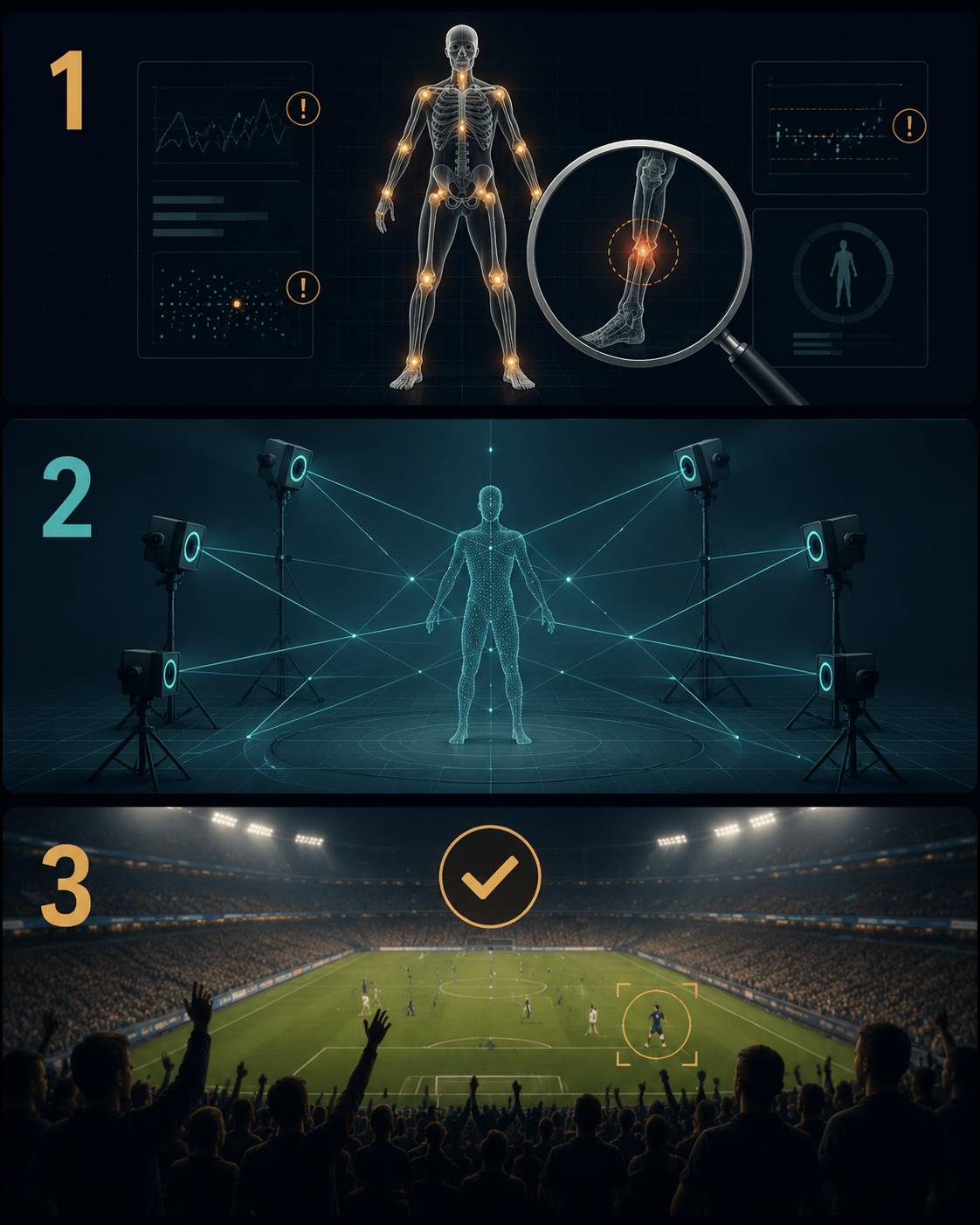
| Validation layer | Primary question | What it can catch |
|---|---|---|
| Data integrity verification | Are the inputs physically and temporally coherent? | Impossible skeletons, distorted limb lengths, latency problems, calibration failures |
| Gold-standard ground-truth testing | How close is the system to an independent reference standard? | Measurement error that looks plausible until compared with a superior reference |
| Staged live trials | Does the system work under operational pressure? | Workflow friction, live-environment instability, review delays, unexpected deployment constraints |
For medical imaging AI, this first layer should be boring in the best possible way. It should ask whether DICOM ingestion is consistent, whether reconstruction differences are tracked, whether scanner and protocol metadata are preserved, whether annotations line up with anatomy, whether image quality filters behave as intended, and whether preprocessing creates silent distortions. None of that proves clinical benefit. It prevents the model from being built on nonsense.
Ground truth had to come from outside the production system
The second SAOT validation layer moved from input coherence to measurement accuracy. Victoria University’s TRACK program used a 44-camera Vicon motion-capture system with sub-millimeter precision to evaluate SAOT limb tracking against an independent gold-standard reference.[2] This is the layer that turns “the skeleton looks plausible” into “the measured position agrees with a better measurement system.”
That independence is the point. A production system cannot validate its own geometry simply by being internally consistent. It needs a reference standard that is not produced by the same cameras, the same calibration chain, or the same algorithmic assumptions. In imaging AI, the analogue is not merely having labels. It is having a ground-truth process strong enough to carry the claim being made: expert annotation protocols, adjudication rules, inter-rater reliability where appropriate, and a clear account of whether the reference is pathology, radiology interpretation, surgical finding, longitudinal outcome, or another accepted standard.
This is also where production-grade and research-grade claims start to separate. FIFA technical materials report SAOT production performance above 98% offside-specific accuracy with a spatial tolerance below 3 cm, but those figures should be read as FIFA and vendor-reported production claims rather than independently peer-reviewed publication of the full production system.[3] That does not make them useless. It does mean they should not be treated as the same kind of evidence as an independently published study.
The difference becomes clearer beside the independently published YOLOv8 work. A 2026 Scientific Reports study reported 83% accuracy for an offside-detection proof of concept using real broadcast footage, with human assistant referees cited at about 94% accuracy in 1,247 decisions and a 95% confidence interval of 92% to 96%.[4] The same study reported annotation agreement with Cohen’s kappa of 0.87.[4]
Those numbers do not show that YOLOv8 is “worse SAOT.” They show something more clinically relevant: readers can easily see the same sport, the same offside task, and the same computer-vision vocabulary, then glide past the difference between a proof-of-concept and a deployed, heavily instrumented production system. The YOLOv8 study explicitly positioned its approach for sub-elite competitive levels without VAR access, not for autonomous top-tier officiating.[4]
That distinction should feel familiar in clinical AI. A model trained and tested in a well-curated academic dataset is not automatically the same object as a monitored clinical deployment across scanner vendors, technologist habits, patient mix, network constraints, and reporting workflows. A retrospective benchmark may be a legitimate research result and still be inadequate as a deployment argument.
The live layer tested the system after the lab had done its work
SAOT then went into staged live-match trials before World Cup authorization, including tests at the FIFA Arab Cup 2021 and FIFA Club World Cup 2021.[3] This layer did not replace the earlier validation layers. It asked a different question: after data integrity and ground-truth accuracy have been addressed, what happens when the system is inserted into the live environment where timing, review workflow, human oversight, and operational pressure all matter?
Healthcare has a direct parallel here, although the endpoints differ. A radiology AI tool may perform well on a retrospective test set and still fail locally because studies arrive with unexpected protocol variants, because priors are unavailable, because triage flags interrupt reading patterns, because false positives accumulate at the wrong point in the day, or because clinicians do not know when the model is outside its validated operating conditions. Live validation is where those problems become visible.
The SAOT staging also keeps the human role visible. The system was semi-automated, not autonomous; its output entered an officiating process. Medical imaging AI needs the same specificity. A detection model that queues suspected hemorrhage, a segmentation tool that drafts tumor volumes, and a diagnostic classifier that influences a report are not the same operational intervention. Each creates different review burdens, different failure modes, and different consequences when wrong.
Infrastructure is part of the validation claim
One reason production SAOT and a single-camera research prototype should not be casually compared is that they are not buying the same evidence. The production-grade system’s reported performance depended on substantial stadium infrastructure, with estimated investment of $2 million to $4 million per stadium.[3] Premier League materials for its SAOT deployment describe 30 cameras operating at up to 100 frames per second and 10,000 surface mesh points per player.[5] That is a different validation environment from a proof-of-concept operating on broadcast footage.
The clinical version is uncomfortable but necessary. An AI model validated in a tertiary academic center with harmonized scanners, experienced subspecialty annotators, strong informatics support, and continuous monitoring may not transfer cleanly to a community hospital with older equipment, thinner staffing, noisier metadata, and fewer resources for post-deployment surveillance. That is not a moral failure by the community site. It is a deployment condition that belongs inside the validation argument.
For 3D surgical reconstruction, multi-camera calibration problems may look closer to SAOT’s triangulation problem. For radiology models, the camera count becomes scanner variation, acquisition protocol, reconstruction kernel, contrast timing, slice thickness, compression, and archive behavior. The form changes. The validation burden does not.
What medical imaging AI should borrow
The transferable structure is not soccer-specific. It is a discipline of separation. Each validation layer should be able to fail independently, and each should catch errors the others are poorly designed to see.
- Before training or local tuning, inspect data fidelity directly: image orientation, metadata completeness, preprocessing outputs, segmentation plausibility, scanner and protocol distribution, and temporal or routing delays.
- Before making performance claims, define a ground-truth process that matches the claim: independent expert review, adjudication, pathology confirmation, outcome linkage, or another defensible reference standard.
- Before routine clinical reliance, run staged live trials that measure workflow impact, alert burden, failure escalation, clinician override behavior, and model behavior under local operating conditions.
- Before comparing products or publications, separate research prototypes, vendor-reported production claims, peer-reviewed validation studies, and independently audited deployed performance.
This structure also changes how weak evidence should be read. A model with a polished retrospective result but no input-fidelity audit has not finished the first layer. A model with plausible overlays but no independent ground truth has not finished the second. A model with strong test-set performance but no staged local trial has not finished the third. The missing layer tells the evaluator what kind of uncertainty remains.
SAOT does not prove that a medical imaging AI tool is safe, effective, or clinically useful. It offers a defensible testing architecture from another high-stakes computer-vision environment. If a sports officiating system needed separate checks for data integrity, independent ground-truth accuracy, and live operational performance before deployment, medical imaging AI should not be accepted on a single retrospective benchmark and a polished accuracy number.
References
- Heads in the Game, MIT Technology Review, June 2026.
- VU research features in new World Cup offside technology, Victoria University.
- FIFA Inside FIFA technical explainer and media releases, FIFA.
- Nature Scientific Reports 2026 offside-detection YOLOv8 study, Scientific Reports, 2026.
- Premier League and Genius Sports semi-automated offside technology press materials, Premier League and Genius Sports.
Comments
Join the discussion with an anonymous comment.