
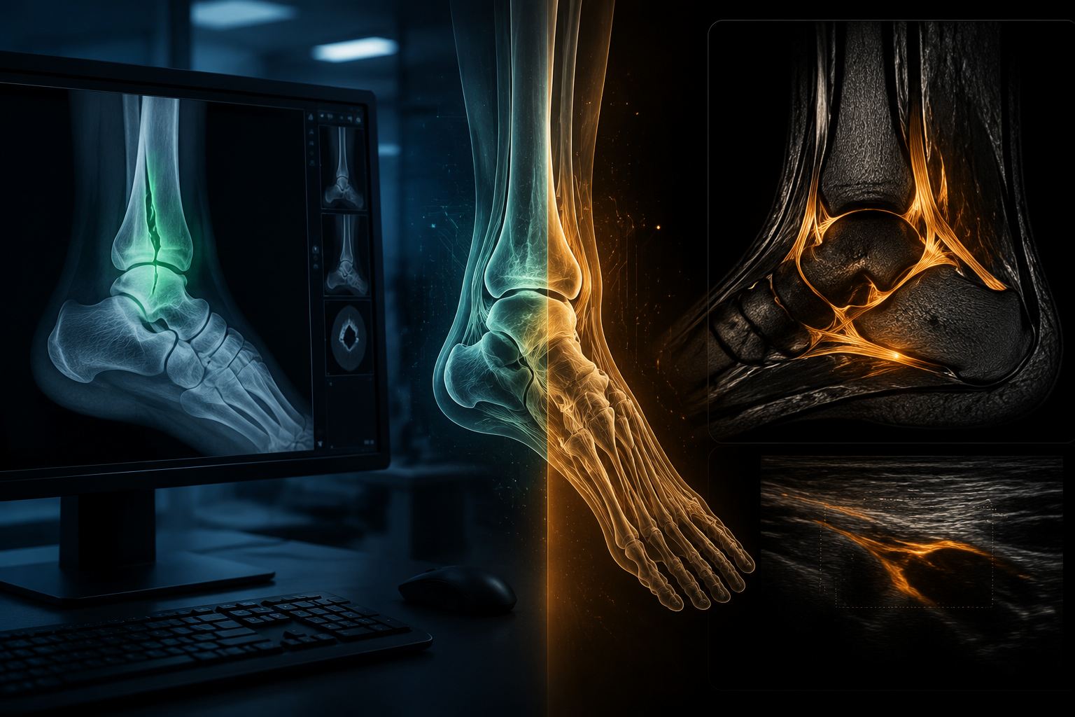
An athlete with a swollen ankle does not arrive with one imaging problem. The first fork is usually plain enough: is there a fracture on radiographs, or is the real concern a ligament, tendon, or instability pattern that needs MRI or ultrasound? That split matters more than any broad claim about AI in sports ankle injury diagnosis, because the evidence is not evenly distributed across those tasks.
The clinical pressure is real. Ankle sprains account for 15–20% of all sports injuries, and recurrent problems are reported in about 70% of patients after ankle sprain.[1] But high clinical volume does not make every AI model equally ready. Fracture detection on X-ray has a much stronger pathway from study performance to deployable clinical decision support. Ligament and tendon assessment on MRI or ultrasound is producing striking research results, but most of that work has not yet cleared the harder tests of external validation, prospective use, and transportability across scanners, operators, and patient populations.
| Imaging task | Current clinical-readiness judgment | Why it sits there |
|---|---|---|
| Ankle and foot fracture detection on X-ray | Deployable as decision support in appropriate workflows | High reported AUCs, external validation evidence, reader-assistance data, and FDA-cleared commercial tools covering ankle or foot imaging.[2][3][4][5] |
| Ligament instability and tendon pathology on ultrasound or MRI | Promising, but not broadly adoption-ready | Very high reported accuracy in selected studies, but limited external validation and little prospective real-world deployment evidence.[6][7] |
| Biomechanics, injury prediction, chatbot triage | Adjacent, not the main diagnostic imaging claim | Useful research areas, but they answer different questions from radiographic, ultrasound, or MRI diagnosis.[8] |
Where AI is strongest: ankle fracture detection on X-ray
Radiographic fracture detection is the part of ankle injury AI where confidence is currently most defensible. The task is visually constrained compared with full soft-tissue interpretation: identify and localize a fracture on a radiograph, then help the reader avoid a miss on a busy list. That does not make the problem easy, especially around subtle, nondisplaced, or overlapping anatomy, but it does make the clinical objective clearer than asking a model to adjudicate chronic instability or tendon degeneration.
The best ankle fracture papers are not merely reporting attractive internal metrics. Ashkani-Esfahani et al. reported an AUC of 0.99 for detecting ankle fractures using deep learning algorithms.[2] Prijs et al. moved the evidence tier higher by developing and externally validating automated ankle fracture detection, classification, and localization, with an AUC of 0.92.[3] That distinction matters. A model can look excellent when tested on data that resemble its development set; it becomes more clinically interesting when it is evaluated outside that immediate environment.
Reader-assistance evidence is equally important because fracture AI is usually not being proposed as a freestanding diagnostician. Guermazi et al. found that AI support improved radiographic fracture recognition sensitivity by 10–20%.[4] In practice, that kind of result is easier to translate into workflow than a model-only metric. It asks whether radiologists or clinicians read differently with AI assistance, whether missed fractures decrease, and whether the tool can support rather than replace ordinary professional judgment.
This is also where regulatory and commercial status changes the conversation. Commercial fracture detection systems catalogued by Kwee and Kwee include Gleamer BoneView, AZmed Rayvolve, and Imagen FractureDetect, with ankle or foot among the covered body regions.[5] The review describes published evidence around commercially available fracture AI, including improved radiologist efficiency and reduced missed fractures.[5] That is not the same as saying every installation will perform identically or that every emergency department should flip a switch tomorrow. It does mean fracture AI has crossed a line that many ankle ligament and tendon models have not: it exists as cleared, usable software with peer-reviewed support, not only as a promising algorithm in a methods paper.
The validation gap is not a footnote
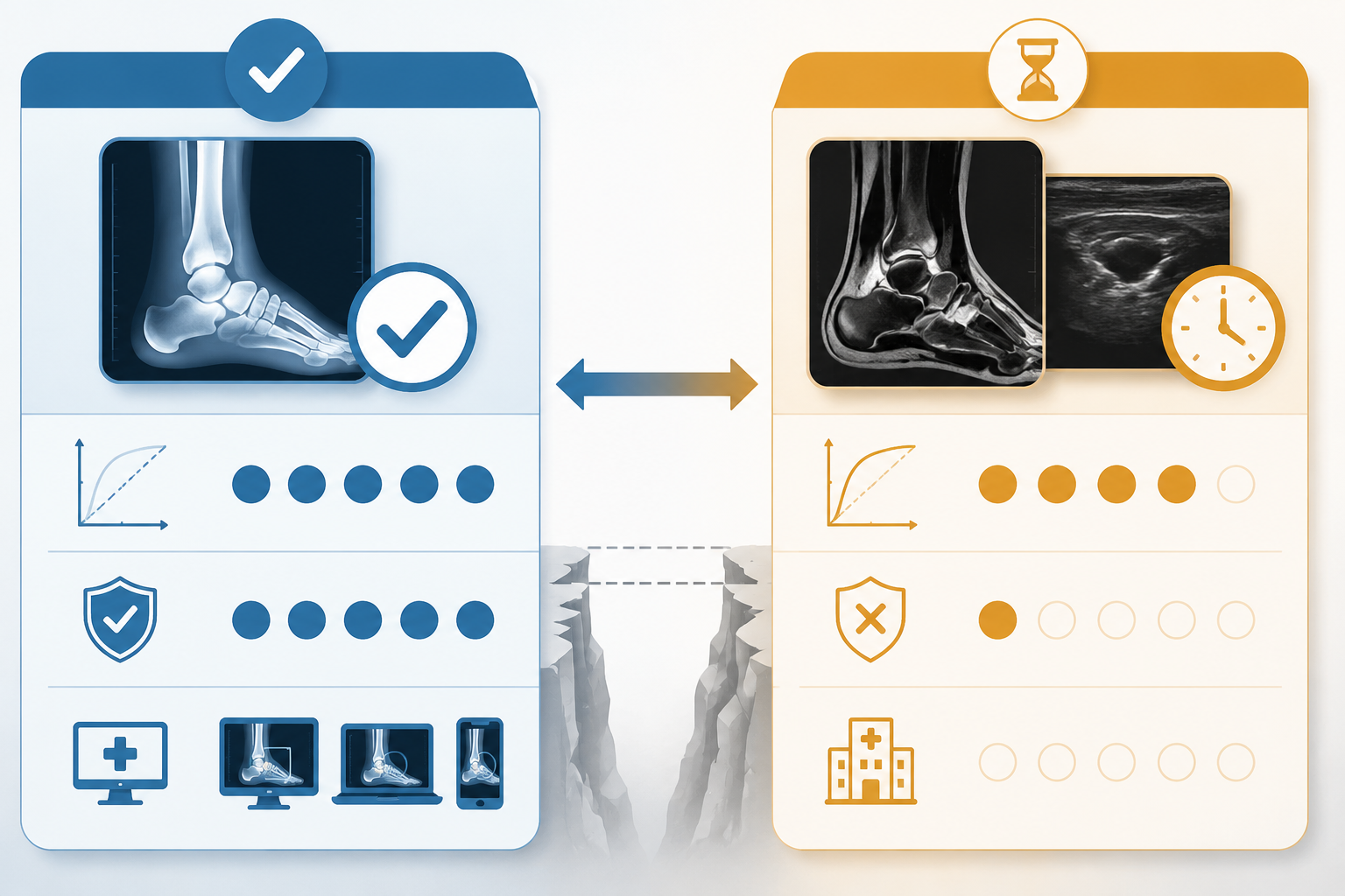
The most uncomfortable number in foot and ankle AI is not a performance score. It is the external validation rate. Gupta et al. reviewed 31 AI studies in foot and ankle surgery and found that only 2 studies, or 6.45%, performed external validation.[6] Nwachukwu et al. also emphasized the same limitation in current foot and ankle AI evidence.[1]
That single fact should change how clinicians read almost every high-performing model in this space. External validation is not academic housekeeping. It is the step that begins to answer whether a model trained in one setting can survive different imaging protocols, equipment, patient mixes, labeling habits, disease prevalence, and referral patterns. In sports ankle imaging, those differences are not trivial. A model trained on selected cases from one institution may encounter a different spectrum in an urgent-care fracture pathway, an elite sports medicine clinic, or a hospital MRI service receiving chronic instability referrals.
This is why a high AUC in a narrow dataset and a lower AUC with external validation should not be read as competing headlines. They answer different questions. The first says the model learned a task under particular conditions. The second begins to say whether the model travels. For clinical adoption, especially in diagnostic imaging, the second question is the one that keeps the radiologist, orthopaedic surgeon, and health IT team out of trouble.
MRI and ultrasound: impressive numbers, narrower readiness
The soft-tissue side of ankle AI deserves attention, not dismissal. Chronic lateral ankle instability, anterior talofibular ligament injury, Achilles tendinopathy, and Achilles tendon injury classification are clinically consequential. They influence rehabilitation, bracing, return-to-play decisions, injection decisions, surgical referral, and patient expectations. They are also harder AI targets than binary fracture detection on radiographs because imaging appearance may overlap with chronic change, partial injury, degeneration, prior treatment, and operator-dependent acquisition.
Kamachi et al. reported one of the more eye-catching results: three deep learning architectures—VGG16, ResNet50, and EfficientNetV2M—were used on ultrasound images of the anterior talofibular ligament to diagnose chronic lateral ankle instability, reaching 98.9% accuracy and an AUC of 0.998.[7] Those numbers are too strong to wave away as mere hype. They suggest that ultrasound image patterns may contain enough signal for automated classification, at least under the study conditions tested.
The same applies to reported work on Achilles pathology. Foot and ankle AI reviews include ultrasound work by Wang et al. reporting an AUC of 0.99 for Achilles tendinopathy and MRI work by Kapinski et al. reporting 97.6% accuracy with 99.45% specificity for Achilles tendon injury classification.[6][1] For a sports medicine clinician who routinely sees posterior ankle pain, these findings are clinically tempting. They point toward tools that might eventually standardize interpretation, flag subtle abnormalities, or support less experienced readers.
But soft-tissue ankle imaging cannot borrow the readiness status of fracture AI. Ultrasound depends heavily on acquisition technique, probe position, dynamic maneuvers, and operator skill. MRI protocols vary by magnet, coil, sequence, slice thickness, field strength, and institutional preference. Chronic instability is also not a single image finding; it sits between anatomy, symptoms, examination, laxity, recurrent sprain history, and functional impairment. A model that performs well on curated images may still need to prove that it can support a clinician when the scan is imperfect, the patient has previous injury, or the imaging finding does not map neatly onto symptoms.
What would make ligament and tendon AI more clinically persuasive?
For ligament and tendon models, the next evidence step is not another isolated accuracy peak. The more useful studies would test the same model outside its development site, report performance across equipment and operators, include clinically messy cases, and show how the output changes reader behavior. Prospective testing would be especially valuable because it exposes the tool to the ordinary sequence of care: the referral question, image acquisition, preliminary interpretation, specialist review, treatment decision, and follow-up.
A model that identifies chronic lateral ankle instability on selected ultrasound images may be scientifically impressive. A model that helps a sports medicine clinic triage equivocal instability cases, reduces unnecessary MRI, improves confidence in return-to-play planning, or standardizes referrals across operators would be clinically different. The literature is not yet consistently at that second level.
Deployment is a workflow question, not just a model question
In a radiology department or sports medicine network, AI readiness depends on more than model discrimination. Someone has to decide where the output appears, who sees it, whether it changes reporting priority, how false positives are handled, and whether the system is monitored after installation. Fracture AI fits relatively well into that workflow: radiographs are common, turnaround expectations are short, misses have immediate consequences, and the AI result can be presented as a second reader or triage flag.
For ankle X-rays, the practical use case is straightforward. The model can flag a suspected fracture for the interpreting clinician, support prioritization on a crowded list, or act as a safety net before discharge. The human reader still adjudicates the image, integrates the examination, and decides whether additional imaging or referral is needed. That division of labor is why decision-support evidence, such as improved reader sensitivity, carries more clinical weight than a model score alone.[4]
Soft-tissue tools face a less settled workflow. If an ultrasound model suggests anterior talofibular ligament abnormality, does it alter rehabilitation, prompt MRI, support surgical referral, or simply echo what an experienced operator already saw? If an MRI model classifies Achilles tendon injury, does it improve reports from general radiology settings, or only perform well when the cases are already tightly labeled? These questions are answerable, but they require studies designed around clinical use rather than retrospective image classification alone.
What this article is not counting as diagnostic imaging readiness
There is a wider world of AI around ankle sprains: biomechanics, kinematics, kinetics, wearable data, injury-risk prediction, and chatbot-style decision support. Those tools may become useful in prevention, rehabilitation, training load management, or patient navigation. They should not be treated as evidence that AI can diagnose an ankle fracture, ligament tear, or tendon injury on imaging.
Teoh et al. reviewed AI in the kinematics and kinetics of ankle sprains, a related but distinct area from diagnostic image interpretation.[8] That boundary is worth keeping clean. A model predicting injury risk from movement data and a model detecting a fracture on X-ray live in different evidentiary worlds. So does a chatbot vignette compared with a validated imaging algorithm. Each may have value, but none should be used to inflate the readiness of another.
The clinical-readiness judgment
AI in sports ankle injury diagnosis is ready only for some questions. For ankle and foot fracture detection on X-ray, the evidence is strong enough to support use as clinical decision support in appropriate workflows: high AUCs, external validation, reader-assistance data, and cleared commercial products all point in the same direction.[2][3][4][5]
For ligament instability and tendon pathology on MRI or ultrasound, the research is promising and in some cases technically impressive. It is not yet in the same readiness category. Until external validation, prospective testing, and real-world deployment evidence become routine, those models should be read as worth watching closely—not as broadly dependable tools for changing sports ankle imaging workflow.
References
- Artificial intelligence in foot and ankle surgery: current concepts — PMC, 2023
- Detection of ankle fractures using deep learning algorithms — PubMed
- Development and external validation of automated detection, classification, and localization of ankle fractures — PubMed
- Improving radiographic fracture recognition performance and efficiency using artificial intelligence — PubMed
- Commercially available AI tools for fracture detection: the evidence — PMC, 2024
- Advancements in AI for Foot and Ankle Surgery: A Systematic Review — PMC, 2023
- Using deep learning for ultrasound images to diagnose chronic lateral ankle instability — PMC, 2025
- Scoping review of AI in kinematics and kinetics of ankle sprains — ScienceDirect, 2024
Comments
Join the discussion with an anonymous comment.