
Tongue cancer is not mainly a story about diagnostic impossibility. It is a story about a visible, examinable disease that still too often reaches care after the window for easier treatment has narrowed. In SEER data for 2016–2022, only 28% of tongue cancers were diagnosed at the localized stage, while 58% already had regional lymph node involvement at presentation. The survival gradient is blunt: five-year relative survival was 88.2% for localized disease, 69.9% for regional disease, and 38.4% for distant disease; overall five-year relative survival was 71.4%.[1]
That is the early-detection gap behind tongue cancer symptoms that are easy to underestimate. The problem is not that every early lesion announces itself. Early-stage tongue cancer may be painless, and its first visible signs can resemble findings that busy primary care, dental, or general medical settings encounter constantly: a sore that does not heal, a red or white patch, a painful area, or a lesion that seems nonspecific until it persists. The danger is that the early presentation can look clinically ordinary, while the later presentation behaves anything but ordinarily.
This is where artificial intelligence becomes clinically interesting. The useful question is not whether a neural network can produce a high accuracy number on a curated image set. The useful question is whether it changes the odds in the same uncertain space where a general physician sees a tongue lesion, lacks specialist certainty, and must decide whether the finding deserves escalation.
The clinical gap AI is trying to enter
| Disease stage at diagnosis or outcome | SEER finding |
|---|---|
| Localized diagnosis | 28% of tongue cancers, 2016–2022 [1] |
| Regional lymph node involvement | 58% of tongue cancers at presentation [1] |
| Five-year relative survival, localized | 88.2% [1] |
| Five-year relative survival, regional | 69.9% [1] |
| Five-year relative survival, distant | 38.4% [1] |
| Estimated new cases in 2026 | 20,420 [1] |
| Incidence trend | Rising 1.0% annually [1] |
The localized-stage statistic matters because it prevents the discussion from drifting into gadget enthusiasm. If barely more than one in four cases are diagnosed while still localized, the system is already failing many patients before advanced oncology decisions begin. Earlier recognition does not guarantee cure, and a screening algorithm cannot erase referral delays, biopsy access, or treatment disparities. But the stage distribution makes a narrower point that is hard to ignore: there is room for a tool that helps front-line clinicians separate harmless-looking persistence from lesions that need specialist assessment.
For that reason, the most relevant AI evidence is comparative. A model that performs well against other models may be technically interesting. A model that performs against general physicians and approaches specialist judgment is clinically more informative.
The Heo study is the key test case
Heo and colleagues’ 2022 Scientific Reports study is the most clinically meaningful piece of evidence in this set because it does several things that many diagnostic AI papers do not. It uses endoscopic images, draws from more than one hospital, includes external validation, and compares model performance with physicians rather than stopping at internal model metrics.[2]
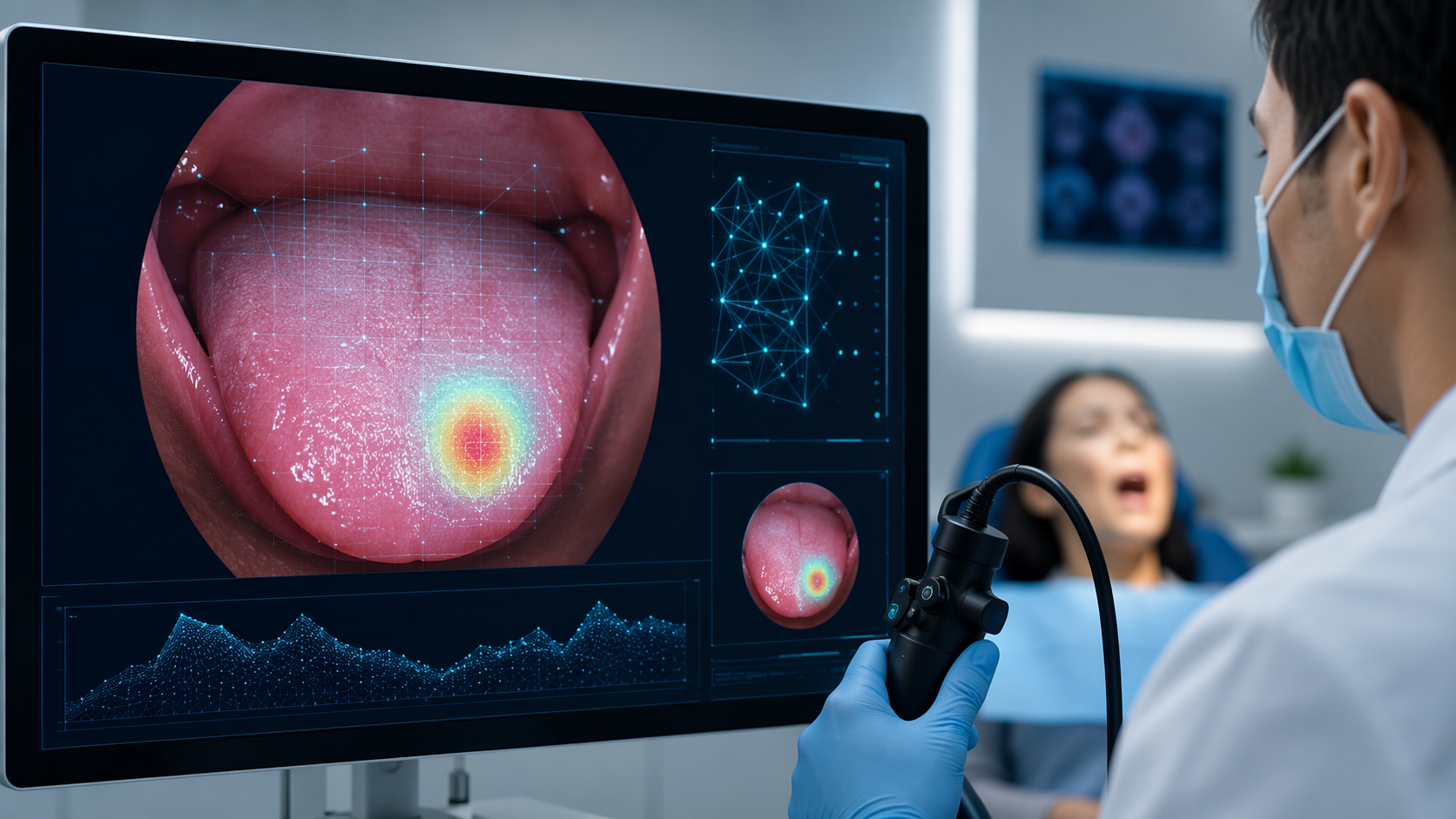
The investigators trained a DenseNet169 model on 5,576 validated endoscopic images collected from five South Korean university hospitals. In external validation, the model achieved an AUROC of 0.895 and an AUPRC of 0.918. It also outperformed general physicians in diagnostic accuracy, 84.7% versus 75.9%, and showed moderate agreement with oncology specialists, with a kappa value of 0.685.[2]
Those details matter more than the architecture name. External validation gives the result some distance from the common failure mode in medical AI: learning the quirks of one dataset and then appearing brilliant inside that dataset. A five-hospital image source does not make the model universal, but it does give the finding more credibility than a one-room retrospective experiment.
The physician comparison is even more important. General physicians are often positioned at the earliest decision point: reassure, observe, refer, photograph, biopsy, or escalate. A model that moves from 75.9% physician accuracy to 84.7% model accuracy in this study is not merely reporting a leaderboard result; it is suggesting a possible role as a second reader in the part of care where missed detection begins.[2]
The kappa result should be read with similar restraint. A kappa of 0.685 indicates moderate agreement with oncology specialists, not replacement of specialist assessment. In practical terms, that is still meaningful. If an AI system can bring generalist interpretation closer to specialist-level judgment, its value may be greatest before the specialist ever sees the patient: during triage, referral justification, and prioritization of suspicious lesions.
There are limits that cannot be brushed aside. The dataset came from South Korean university hospitals. Tongue lesion epidemiology, imaging practices, referral patterns, camera quality, and case mix may differ elsewhere. A model trained and validated in this environment may not behave the same way in a community clinic, dental office, urgent care setting, or health system serving a different population. External validation across hospitals is better than internal validation alone; it is not the same as broad prospective deployment.
Why AUROC, AUPRC, accuracy, and kappa are not interchangeable
Diagnostic AI papers often present a cluster of metrics as if they all say the same thing. They do not. Accuracy tells how often the model is right at a chosen threshold, but it can be distorted when one class dominates the dataset. AUROC describes how well the model separates classes across thresholds. AUPRC is especially relevant when the positive class is less common, because it focuses on precision and recall. Kappa describes agreement beyond chance between readers or systems.
For tongue cancer early detection, the threshold question is not abstract. A conservative threshold may reduce false positives but miss subtle cancers. A sensitive threshold may catch more suspicious lesions but increase referrals and biopsies. The right operating point depends on where the tool is used: front-line triage, specialist support, documentation review, or population screening. The Heo study is useful because it reports discrimination metrics and reader comparison rather than asking the reader to trust a single high percentage.[2]
The wider literature is promising, but structurally weak
The broader AI oral-cancer screening literature supports cautious interest, not clinical triumphalism. A 2026 systematic review in Frontiers in Oncology reviewed 63 studies and found that more than 90% were single-center retrospective designs without external validation. The review also described a trend from 2021 to 2026 toward multimodal fusion, combining imaging with clinical metadata.[3]
That finding is the needed counterweight to the stronger single-study signal. Retrospective single-center work is often where useful models begin, but it is also where models can look cleaner than the clinical reality they are meant to enter. Images may be better framed than routine images. Labels may reflect selected cases rather than the full mess of uncertain mucosal disease. Controls may be easier than real-world differentials. A model may learn site-specific imaging artifacts, lighting, documentation habits, or referral-enriched case mixes.
The multimodal trend is sensible. A tongue image alone may not capture duration, pain, risk factors, prior lesion history, lesion evolution, or clinician concern. Clinical metadata can help, but it can also introduce new problems: missing fields, inconsistent terminology, documentation bias, and workflow burden. A multimodal model that works in a retrospective database still has to prove that clinicians can feed it reliable inputs during an ordinary visit.
The tongue-cancer-specific evidence base is also being consolidated. A 2025 systematic review with meta-analysis focused specifically on artificial intelligence in tongue cancer diagnosis, which is important because much of the surrounding literature is actually oral-cancer literature with tongue cases included as a subset.[4]
That distinction should be preserved. Oral cancer is not interchangeable with tongue cancer for purposes of model performance. A system trained on heterogeneous oral sites may be useful, but its headline performance cannot automatically be treated as tongue-specific performance unless the study reports and validates that subgroup clearly.
Small high-accuracy studies need a slower read
Some newer architecture papers report numbers that look stronger than the evidence structure beneath them. Benil and colleagues reported 97.66% training accuracy using VGG16 on 374 tongue images compiled from public internet sources.[5]
That result may be useful as an engineering experiment, but it should not be mistaken for a deployable clinical estimate. A dataset of 374 images is small for a visual diagnostic task. Public internet images may overrepresent obvious, well-lit, educational, or advanced lesions and may underrepresent the ambiguous findings that create the real early-detection problem. Training accuracy also says much less than external validation. It can show that a model fit the material it saw; it does not show that the model will help a general physician decide what to do with a subtle lesion in clinic.
Liu and Bagi’s 2025 Scientific Reports paper illustrates an even sharper problem. The authors reported 99.54% accuracy using a 19-layer custom CNN on 1,298 lip and tongue images, but the mean AUC on ROC analysis was only 0.6026.[6]
Those two numbers do not tell the same story. Very high accuracy with a weak AUC can occur when class balance, threshold selection, or dataset structure allows a model to be right often without ranking positive and negative cases well across thresholds. In a screening or early-detection context, that is not a minor technical footnote. If the curve underneath the accuracy figure is weak, the model may not be dependable when the threshold changes, the population changes, or the disease prevalence changes.
There is also an anatomical boundary problem. Liu and Bagi studied lip and tongue images, not tongue cancer alone. That does not make the study irrelevant, but it narrows what can be claimed from it. A lip-and-tongue classifier may learn features that do not transfer evenly across sites. The same caution applies to broader oral-cancer models: useful signals, limited tongue-specific certainty.
Where AI could actually help in early detection
The most plausible near-term role is not autonomous screening of everyone with a mouth symptom. It is assisted triage in settings where lesions are already being seen or imaged: primary care, dentistry, otolaryngology intake, oral medicine, oncology follow-up, or teleconsultation workflows. The tool’s job would be to reduce the chance that a persistent sore or patch is normalized too early.
In a realistic workflow, the model would not simply output “cancer” or “not cancer.” It would flag suspicious images, support referral decisions, prompt repeat imaging when quality is poor, or help prioritize specialist review. The consequence of a positive result would still need to be a human clinical action: examination, referral, biopsy consideration, or documented follow-up. The consequence of a negative result would need guardrails, especially when a lesion persists.
This is also where performance against general physicians matters. If a model can improve generalist recognition without pretending to replace specialists, it addresses the point in care where the localized-stage opportunity is most likely to be lost. The Heo study’s comparison suggests that this is no longer a purely speculative use case.[2]
But the deployment bar should remain high. A tool intended for early detection would need prospective testing, predefined thresholds, representative populations, image-quality standards, subgroup reporting, and measurement of downstream effects. It is not enough to know whether the model classifies images correctly. Clinicians and health systems need to know whether it changes referrals, time to biopsy, false reassurance, unnecessary procedures, and ultimately stage at diagnosis.
The current evidence supports a detection aid, not a screening promise
AI for tongue cancer detection has crossed an important credibility threshold. In the best comparative evidence here, a deep learning model trained on multi-hospital endoscopic images outperformed general physicians and approached oncology-specialist agreement. Given the survival difference between localized and distant disease, that kind of assistive performance deserves attention.
The evidence has not crossed the threshold for broad screening claims. Too much of the field remains retrospective, single-center, and thin on external validation. Some high-accuracy reports are weakened by small datasets, training-only results, mixed anatomical sites, or contradictory metrics. The most careful conclusion is also the most useful one: the early-detection gap in tongue cancer is real, AI may help close part of it, and the literature is not yet mature enough to act as if the problem has been solved.
References
- Cancer Stat Facts: Tongue Cancer, SEER
- Deep learning model for tongue cancer diagnosis using endoscopic images, Scientific Reports, 2022
- Artificial intelligence and its application in early oral cancer screening: a systematic review, Frontiers in Oncology, 2026
- Artificial Intelligence in the Diagnosis of Tongue Cancer: A Systematic Review with Meta-Analysis, Biomedicines, 2025
- Detect pre-cancerous tongue lesions for early oral cancer diagnosis using deep learning algorithm, Scientific Reports, 2025
- A tailored deep learning approach for early detection of oral cancer using a 19-layer CNN on clinical lip and tongue images, Scientific Reports, 2025
Comments
Join the discussion with an anonymous comment.