
When Nivea disclosed in July 2026 that she had been diagnosed with leukemia, the public details were few: she is 44, she spoke about the diagnosis on Cadillac Chronicles, the episode aired July 21, she said she had been diagnosed earlier in 2026, and she reported responding well to treatment.[1] That is enough to explain why people searched for “nivea leukemia diagnosis details what we know.” It is not enough to infer her leukemia subtype, prognosis, diagnostic timeline, treatment regimen, or whether any artificial intelligence tool was involved in her care.
That boundary matters. Leukemia is not a single diagnostic label that simply appears on a chart after one abnormal blood count. In real practice, the first abnormality has to be reconciled with morphology, flow cytometry, cytogenetics, and molecular testing before clinicians can confidently name the disease and select the next step. The waiting period can be clinically important and emotionally brutal: a patient may know that something serious is wrong before the team can say precisely what kind of leukemia it is.
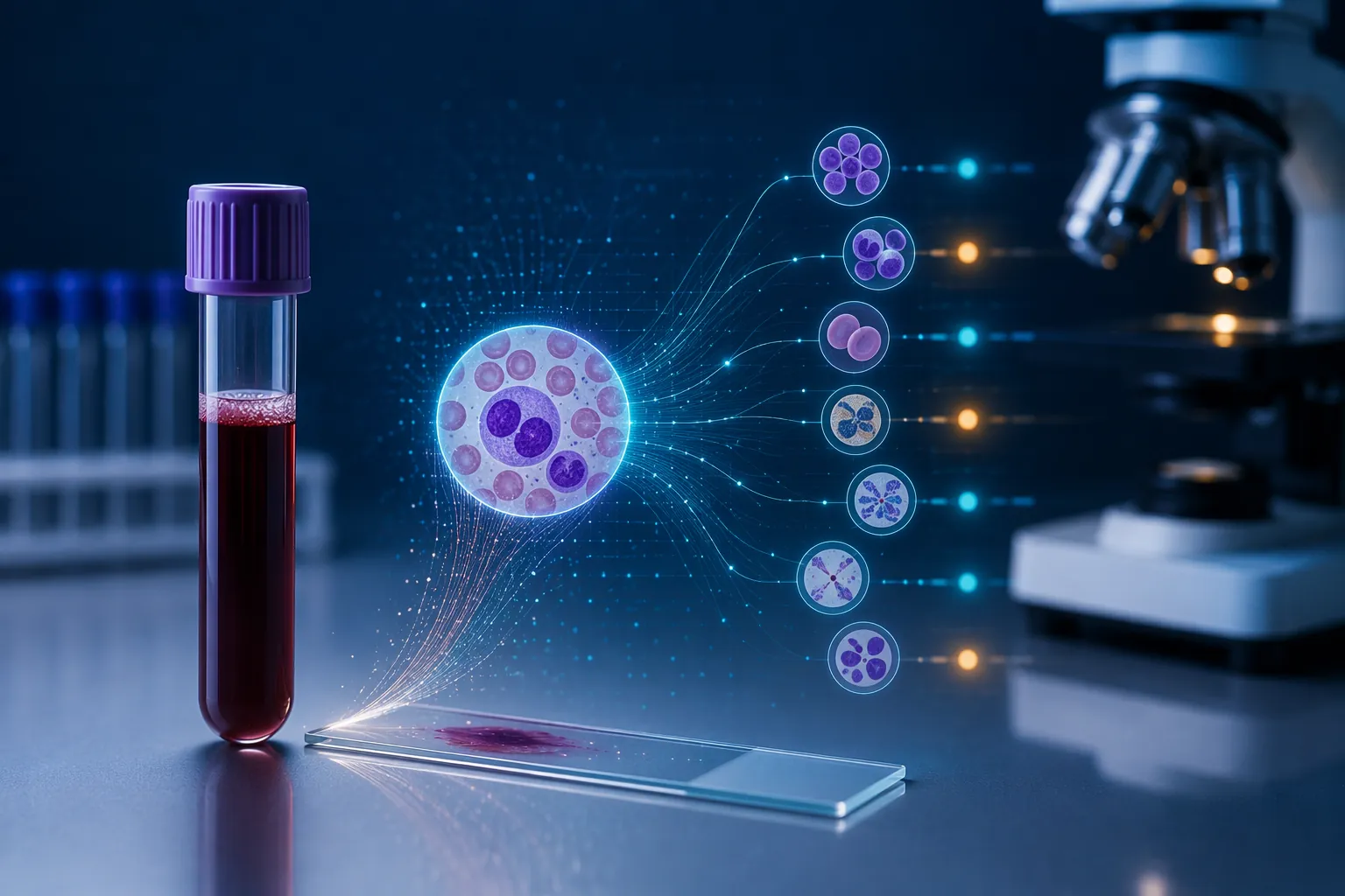
The useful question raised by Nivea’s announcement is therefore not whether AI could explain her case. It cannot, because the public record does not contain the necessary clinical information. The better question is where AI-assisted leukemia diagnostics now fit when a patient enters that uncertain interval between “something is wrong” and “we know the subtype.” The strongest 2025–2026 evidence suggests that AI can sometimes shorten parts of that pathway, but it does not remove the pathway.
The New AI Tools Are Not All Looking at the Same Disease Signal
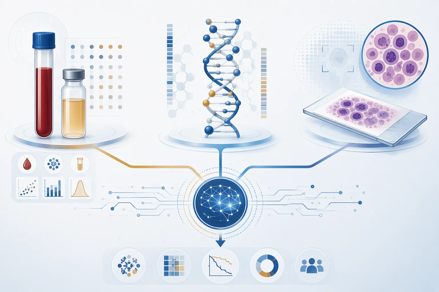
A single phrase like “AI detects leukemia” hides the most important distinction: different systems are reading different inputs and answering different clinical questions. Some use routine laboratory values. Some classify molecular or epigenomic patterns. Some analyze blood-smear images. Those are not interchangeable roles.
| Tool | Main input | Reported clinical role | Key reported performance or timeline |
|---|---|---|---|
| AI-PAL | Routine CBC and coagulation parameters | Screening/classification support for acute leukemia patterns, including AML and APL | AUROC 0.94 for AML and 0.98 for APL with confidence cutoff in international validation; refinement reached 0.84 AUROC with far fewer exclusions |
| MARLIN | DNA methylation analyzed with a neural-network classifier | Acute leukemia subtype classification | Classified 38 subtypes in under 2 hours from biopsy in reported work |
| ALMA | Patient methylation matched to a methylation atlas | AML subtype classification aligned with WHO-defined categories | Matched to 27 WHO-defined subtypes and reduced the diagnostic window to 2–3 days in the reported description |
| cAItomorph | Peripheral blood-smear images | Malignant versus non-malignant triage and support for deciding who needs bone marrow aspiration | 0.97 AUC; could reduce unnecessary bone marrow aspirations by 35% while maintaining 100% sensitivity for acute leukemia in the study |
The table is intentionally uneven because the tools are uneven by design. AI-PAL is attractive because it looks for signal in tests many hospitals already run. MARLIN and ALMA are about faster subtype resolution from molecularly informative data. cAItomorph is closer to the front door of hematopathology triage, where a blood smear may help determine who should undergo an invasive bone marrow procedure.
AI-PAL Shows Both the Promise and the Trap
AI-PAL is the most revealing example because its strengths and limitations sit in the same study. Published in Nature Communications in March 2026, the model used XGBoost and routine complete blood count plus coagulation parameters, then underwent international validation across 6,206 patients at 20 centers in 16 countries on five continents; 43% of participating sites were in low- and middle-income settings.[2]
That design matters because routine blood counts and coagulation results are not exotic inputs. A model that can extract useful leukemia classification signal from those values could be valuable in settings where advanced molecular testing is delayed, centralized, or difficult to access. In the reported validation, AI-PAL achieved an AUROC of 0.94 for acute myeloid leukemia and 0.98 for acute promyelocytic leukemia when a confidence cutoff was applied.[2]
The caution is in the cutoff. The initial high-performance approach excluded 70.8% to 92.5% of patients, depending on the task, because the model did not issue confident classifications for many cases.[2] That is not a small implementation detail. In a live diagnostic workflow, excluded patients still need an answer, and the clinical team still has to decide what to do while waiting.
The authors later refined the approach using isolation forest outlier detection, reducing exclusions to 12.1% while reaching an AUROC of 0.84.[2] That tradeoff is more operationally honest: a system that covers more patients may perform less impressively, while a system with spectacular metrics may be speaking only about a selected subset. Both numbers are useful, but they answer different questions.
The site-level variability is even more important. AI-PAL’s APL performance was reported as AUROC 0.98 in Spain but 0.63 in Madagascar, with differences attributed to feature distributions and missing coagulation data.[2] That is the line that should stop any universal claim in its tracks. A leukemia AI model is not validated “globally” just because it has crossed borders; it has to survive the data patterns, missingness, instruments, referral behavior, and laboratory practices of the places where it will actually be used.
MARLIN and ALMA Compress the Subtype Wait
If AI-PAL is compelling because it starts with ordinary lab data, MARLIN and ALMA are compelling because they target the classification bottleneck. Acute leukemia care depends heavily on subtype. The sooner a team can align morphology with molecular classification, the sooner it can move from suspicion to a more specific diagnostic and therapeutic plan.
MARLIN, described by the Broad Institute and Dana-Farber in 2025, uses DNA methylation data with a neural-network classifier to classify acute leukemia into 38 subtypes in under 2 hours from biopsy material.[3][4] The tool was trained on more than 2,500 samples and was reported to resolve cryptic DUX4 rearrangements that conventional methods can miss.[3][4]
That is not merely a faster label printer. Cryptic rearrangements are exactly the kind of finding that can be hard to reconcile when routine tests do not line up cleanly. A classifier that helps surface such patterns faster could reduce the time a patient and clinician spend with an incomplete subtype picture. The evidence supports that narrower claim; it does not support replacing the diagnostic team.
ALMA, reported by UF Health in 2025, uses a methylation atlas approach, matching patient methylation patterns to 27 WHO-defined acute myeloid leukemia subtypes across 3,300 samples.[5] UF Health described the workflow as reducing diagnosis from weeks to 2–3 days using a laptop-sized sequencer.[5] That time difference is clinically meaningful when the alternative is waiting for multiple specialized results to come back in sequence.
MARLIN and ALMA also show why “AI accuracy” is too blunt a phrase. The clinically relevant question is not only whether a model is correct in aggregate. It is whether the input can be obtained quickly enough, whether the classifier recognizes the relevant subtype, whether the output integrates with WHO-defined categories and local reporting practice, and whether discordant findings trigger the right review rather than being smoothed over by a confident software result.
The Blood Smear Is Becoming More Computable
Peripheral blood morphology has always carried information, but its interpretation depends on expertise, sample quality, and the specific clinical question. cAItomorph enters at that point. Published in Leukemia in March 2026, the transformer-based model analyzed peripheral blood smears and achieved a 0.97 AUC for malignant versus non-malignant classification.[6]
The more patient-facing result is not the AUC by itself. In the reported study, cAItomorph could reduce unnecessary bone marrow aspirations by 35% while maintaining 100% sensitivity for acute leukemia.[6] If confirmed prospectively, that would matter because bone marrow aspiration is not an abstract diagnostic step. It is a procedure patients feel, schedule around, and sometimes undergo while clinicians are still sorting out how likely an acute leukemia diagnosis really is.
The phrase “while maintaining sensitivity” is doing heavy work. A triage tool that misses acute leukemia is not acceptable simply because it reduces procedures. A triage tool that helps spare some patients from unnecessary aspiration while still catching acute leukemia would be different. The study result is promising, but the practical question is whether that performance holds when the model encounters routine variation in smear preparation, scanner hardware, staining, referral mix, and borderline cases.
A 2025 systematic review and meta-analysis of AI detection of AML from blood images gives useful calibration. It reported random-effects accuracy of 0.956 and sensitivity of 0.858, while also emphasizing high heterogeneity across study designs.[7] That combination should feel familiar by now: image-based AI can perform strongly, but pooled performance does not erase differences in data source, labeling quality, model architecture, and validation design.
Accuracy Is Not the Same as Clinical Permission
The 94% to 98% range in recent leukemia AI reports is real enough to take seriously, but it is not the same thing as a standalone diagnostic claim. Study performance is produced under defined inclusion criteria, available data fields, selected endpoints, and retrospective or controlled validation conditions. A hospital workflow is messier. Patients arrive with missing values, partial workups, transfusions, infections, anticoagulation, prior treatment, and samples that do not behave like the training set.
As of July 2026, the evidence supplied for these tools does not establish that any of them is FDA-approved, or equivalently approved, as a standalone leukemia diagnostic. Nor does it establish that prospective trials have validated them in live clinical workflows. That distinction is not regulatory pedantry. It determines who is accountable when a model is wrong, when it abstains, when it conflicts with morphology, or when it performs well in one institution and poorly in another.
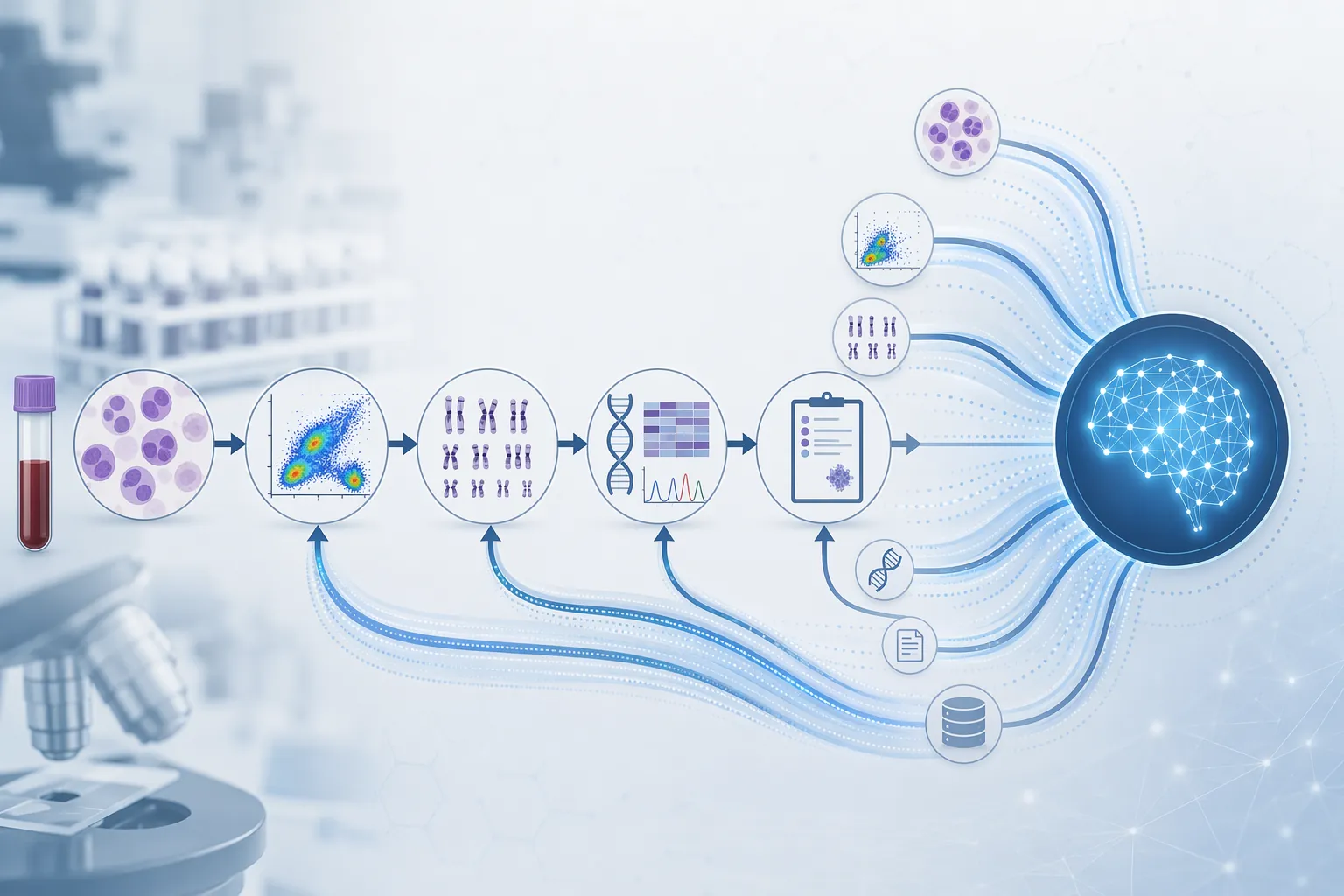
The safest current reading is that these systems are accelerators and classifiers inside a diagnostic pathway. AI-PAL may help flag patterns in routine blood and coagulation data. MARLIN and ALMA may shorten the wait for molecularly meaningful subtype information. cAItomorph may help triage blood-smear findings and reduce unnecessary marrow procedures if its reported sensitivity holds in prospective use. None of that makes morphology, flow cytometry, cytogenetics, or molecular testing old-fashioned friction.
The clinician still has to integrate the result. A model may produce a probability; the hematopathologist has to decide whether the smear, immunophenotype, karyotype, fusion testing, sequencing, and clinical presentation form a coherent diagnosis. When they do not, the discordance is not a nuisance to be averaged away. It may be the most important part of the case.
What This Means for Patients Waiting for a Name
For a patient newly told they have leukemia, the promise of AI is not that a computer independently “finds cancer” and ends the diagnostic process. The more meaningful promise is narrower: fewer days spent waiting for subtype information, fewer avoidable procedures, faster recognition of patterns that need urgent review, and earlier alignment among the tests that already define modern leukemia diagnosis.
Nivea’s announcement belongs first in that human frame. Publicly, we know she disclosed a leukemia diagnosis, said it came earlier in 2026, and reported responding well to treatment.[1] We do not know the subtype or diagnostic pathway. Her disclosure should not be used to imply that any specific AI system would have changed her care.
What her story does make visible is the rupture that comes before classification: a person learns they have a blood cancer, while the clinical system works to name it accurately enough to treat it. As of Q3 2026, AI-assisted leukemia diagnostics are becoming credible tools for shortening and sharpening parts of that work. They still need prospective testing, appropriate regulation, transparent failure handling, and proof across the settings where patients actually receive care.
References
- Nivea, 44, Reveals Cancer Diagnosis: 'I Live in Gratitude', People, July 22, 2026.
- AI-PAL: artificial intelligence for the diagnosis of acute promyelocytic leukemia using routine laboratory data, Nature Communications, March 2026.
- New AI-based diagnostic tool uses epigenomics to accelerate acute leukemia diagnosis, Broad Institute.
- New diagnostic tool developed at Dana-Farber revolutionizes acute leukemia diagnosis, Dana-Farber Cancer Institute, 2025.
- AI tool speeds acute myeloid leukemia diagnosis, UF Health College of Pharmacy, August 4, 2025.
- cAItomorph: a transformer-based artificial intelligence model for the diagnosis of hematological malignancies in peripheral blood smears, Leukemia, March 2026.
- Artificial intelligence for acute myeloid leukemia detection from blood cell images: a systematic review and meta-analysis, Frontiers in Big Data, 2025.
Comments
Join the discussion with an anonymous comment.